Cytology and histology - what is the difference?
The difference between cytological analysis and histological examination is that cells are studied, not tissue sections. This means that final conclusions are drawn on the basis of changes that have occurred in the nucleus, cytoplasm, nuclear-cytoplasmic ratio, formation of complexes and cell structures.
Many people have no idea what such a procedure is, since the technique differs significantly from others. Much is different here in that each organ has its own technique, so a smear, section method, and tissue films can be used here.
It is very important to adhere to the approved algorithm; all research rules must be strictly observed. When a tissue fragment is obtained, it should be placed in ethanol (formalin can be used), then a section is made (it should be thin) and it must be stained, special substances are used for this.
There are several ways to stain thin sections. The most commonly used is eosin, and hematoxylin is also widely used. Such substances affect the cut tissue, causing its color to change.
If hematoxylin is used for staining, the tissue turns blue and the proteins turn red. After all the manipulations, the finished sample must be examined using an electron microscope to identify dangerous cells that can cause illness.
There is another way, when tissue sections are placed in special paraffin; balm can also be used for this purpose. Various microscopes are used to conduct research; if you use a phase-contrast microscope, you can examine samples that cannot be examined with conventional microscopes.
The difference between cytological analysis and histological examination is that cells are studied, not tissue sections. This means that final conclusions are drawn on the basis of changes that have occurred in the nucleus, cytoplasm, nuclear-cytoplasmic ratio, formation of complexes and cell structures.
Where else are these methods used?
In medicine there is the science of embryology. She studies the features of the formation and development of embryos from the moment of conception. New living organisms first consist of one fertilized cell, which is then actively divided into a huge number of them.
To study these processes fully, cytology and histologists are used. The second method, however, due to its traumatic nature, is practically not used on viable embryos. After all, in this case there is a risk of seriously harming them.
But cytology allowed embryologists to learn how to do in vitro fertilization, which gave many childless couples a chance to become parents. Before carrying out this procedure, doctors carefully study all reproductive material in order to select the most viable germ cells from it. This is how cell biology works in practice. Cytology and histology are its main methods, which help not only restore health to a person, but also the birth of a child.
When is a cytology test done?
Cytological examination is used for:
- Preventive examination (screening) Clarification or establishment of a diagnosis of a disease Clarification or establishment of a diagnosis during surgery Monitoring during and after treatment Observation of the dynamics of the process or for early detection of pathological changes
- Preventive examination (screening)
- Clarifying or establishing a diagnosis of a disease
- Clarifying or establishing a diagnosis during surgery
- Control during and after treatment
- Monitoring the dynamics of the process or for early detection of pathological changes
READ MORE: Can there be errors in cytology?
What materials are used for analysis
These can be liquid samples:
- urine, sputum or prostate juice cerebrospinal and amniotic fluid swabs from various organs taken during endoscopy smears of the cervix and smears of the uterine cavity (cytological examination of smears, cytological examination of the cervix) discharge from the mammary glands scrapings and impressions from eroded or ulcerated surfaces, fistulas or wounds fluid from articular and serous cavities
These include materials obtained using aspiration diagnostic puncture, which is carried out using a special thin needle.
In this case we are talking about impressions from removed tissue, such as from a fresh cut surface of tissue removed during surgery or taken for further histological examination.
The materials for further cytological examination are:
- Biological fluids. It is rarely used to diagnose cervical cancer. Such samples are obtained as a result of prostate massage (juice is released from the urethra), and treatment of internal organs during diagnostic minimally invasive operations. To determine pathologies of the respiratory tract, sputum is taken for analysis. In some cases, urine testing may be possible.
- Points. The material is obtained as a result of a diagnostic puncture, for which appropriate needles are used. Depending on the indications, articular fluid, spinal fluid, amniotic fluid in pregnant women, neoplasm cells, muscle tissue of internal organs, and the membranes of the heart are collected.
- Fingerprints and scrapings. In this case, biological material is obtained by applying a glass slide or scraping sections of tissue from an open wound during surgery, the cervix during a biological examination, ulcers, fistulas.
Doctors recommend making an appointment for a test 1 to 2 days after the cessation of menstruation. But to obtain reliable results for the examination, it is necessary to properly prepare:
- 2-3 days beforehand, do not douche the vagina, but simply wash yourself using appropriate intimate hygiene products;
- three days before the examination, stop using various drugs in the form of ointments, suppositories, tampons, spermicides intended for insertion into the vagina;
- 3 - 4 days before the analysis, you must strictly abstain from sexual contact;
- 2 - 3 hours before your visit to the doctor, do not go to the toilet.
The collection of material for analysis is carried out as follows:
- The woman is asked to move to the gynecological chair.
- The doctor uses a dilator to provide access to the cervix.
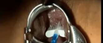
- Using a special instrument that looks like a small brush, the doctor scrapes the surface of the cervix and cervical canal. In some cases (according to indications), the doctor uses an aspirator designed to obtain mucus from the cervical canal.
- The resulting sample is placed in a special tube and labeled accordingly.
READ MORE: What is the cytological picture of a colloid thyroid nodule - LiveAcademy
Often, in the process of taking material for a cytological examination, the gynecologist also makes a smear to analyze the composition of the mixed bacterial flora of the vagina and identify the causative agents of sexually transmitted infections.
Currently, to facilitate the procedure for taking samples and interpreting the obtained data, gynecologists use ready-made test systems ThinPrep PAP Test or SurePath PAP Test.
The use of the latter is recommended by the American FDA, since in clinical studies it showed more reliable results.
Liquids
- urine, sputum, or prostate juice
- cerebrospinal and amniotic fluid
- swabs from various organs taken during endoscopy
- smears of the cervix and smears of the uterine cavity (cytological examination of smears, cytological examination of the cervix)
- discharge from the mammary glands
- scrapings and impressions from eroded or ulcerated surfaces, fistulas or wounds
- fluid from joint and serous cavities
Points
These include materials obtained using aspiration diagnostic puncture, which is carried out using a special thin needle.
Prints
In this case we are talking about impressions from removed tissue, such as from a fresh cut surface of tissue removed during surgery or taken for further histological examination.
Definition
Histology is a discipline devoted to the study of tissues of various organisms, including humans. This is also the name of the process of researching said biological material.
Cytology is the science of what forms the basis of the structure of all living things, that is, of cells. The same word means a method that involves the study of similar structural units within the laboratory.
Purpose of cytological examination
The main purpose of the cytological research method is to obtain an answer to the question of the absence or presence of a malignant neoplasm in the patient whose material was studied.
This method allows you to more accurately determine the nature of the pathological process (benign and malignant tumors), the nature of inflammatory, proliferative, reactive and precancerous lesions.
It is the detailed morphological characteristics of the detected neoplasm that makes it possible to choose the most reasonable method of treatment. be it surgical removal of the tumor, radiation therapy, chemotherapy or a combination of these, depending on the structure of the tumor, its origin, the degree of atypia of its cells and the possible response to treatment.
Cytological examination, compared to other methods, has undoubted advantages in identifying the initial stages of cancer. This research method is used in the diagnosis of tumors in almost any tissue and any organ of the human body.
Thanks to cytological examination, it is possible to detect stomach cancer, bladder cancer, lung cancer, and other organs even in the absence of radiological, clinical and endoscopic manifestations and signs.
The main purpose of the cytological research method is to obtain an answer to the question of the absence or presence of a malignant neoplasm in the patient whose material was studied.
This method allows you to more accurately determine the nature of the pathological process (benign and malignant tumors), the nature of inflammatory, proliferative, reactive and precancerous lesions.
It is the detailed morphological characteristics of the detected neoplasm that makes it possible to choose the most reasonable method of treatment. be it surgical removal of the tumor, radiation therapy, chemotherapy or a combination of these, depending on the structure of the tumor, its origin, the degree of atypia of its cells and the possible response to treatment.
READ MORE: Red blood cells in a smear are normal in women
Cytological examination, compared to other methods, has undoubted advantages in identifying the initial stages of cancer. This research method is used in the diagnosis of tumors in almost any tissue and any organ of the human body.
Thanks to cytological examination, it is possible to detect stomach cancer, bladder cancer, lung cancer, and other organs even in the absence of radiological, clinical and endoscopic manifestations and signs.
Summary
Now you know the difference between cytology and histology. Of course, you won’t be able to read your health status from laboratory preparations, but when you are referred to a particular diagnostic method, you will know exactly what it is.
In medical universities, absolutely all students study the basics of cytology and histology. This gives them the opportunity to understand in more detail the structural features of the human body. Later, some of them decide to go work in the laboratory. To do this, future scientists need to study in detail every nuance and phenomenon that is currently open and studied.
The main thing is to always remember that the human body is a very fragile system, a disruption in the functioning of which can begin with one incorrectly divided cell. Therefore, never neglect preventive examinations and always take all necessary tests.
» data-medium-file=»https://i2.wp.com/unclinic.ru/wp-content/uploads/2019/04/laboratornye-issledovanija.jpg?fit=900%2C600&ssl=1″ data-large- file=”https://i2.wp.com/unclinic.ru/wp-content/uploads/2019/04/laboratornye-issledovanija.jpg?fit=900%2C600&ssl=1″ data-lazy-srcset=”https: //i2.wp.com/unclinic.ru/wp-content/uploads/2019/04/laboratornye-issledovanija.jpg?w=900&ssl=1 900w, https://i2.wp.com/unclinic.ru/wp -content/uploads/2019/04/laboratornye-issledovanija.jpg?resize=768%2C512&ssl=1 768w" data-lazy-sizes="(max-w />
Histological and cytological diagnosis are based on the study of cell samples, but they have fundamental differences. A biopsy is not a test at all. This is the name for taking samples for subsequent microscopic examination. Therefore, all three concepts are not synonymous, as some patients think.


