How the method works
The name of the method combines three branches - immunology, histology and chemistry. Chemicals are used for analysis. The reagents are loaded with antibodies that target cancer antigens. An antibody is a protein, its structure attracts antigen. The protein is stained with enzymes and dyes, which appear when the reaction is positive.
The theoretical foundations of immunohistochemistry were laid in the 40s of the 20th century. Fluorescence was used to detect antigens in frozen cells. Then enzyme-stained antibodies were used. The correspondence between the color reaction and the type of cancer cells was established. Hormones and their receptors were also identified.
Practical application of IHC methods:
- Determination of cell type;
- Study of secretory intracellular processes and features of cellular synthesis;
- Determination of hormones and hormonal receptors.
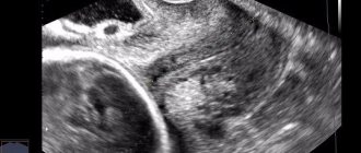
Cells under a microscope after immunohistochemistry
Types:
- Direct method - labeled antibodies are combined with the desired antigens.
- Indirect, “antigen-antibody” - unmarked antibodies find antigens, then their compounds are detected by secondary marked antibodies.
The color of the reaction indicates the tumor status.
What is immunohistochemistry
Immunohistochemistry (IHC) is a method of localizing specific antigens in tissues, based on the recognition of the antigen by the corresponding antibody and identifying the results of this binding at the light-optical level.
The history of IHC dates back to the 1940s when Coons et al developed immunofluorescence techniques to detect antigens in frozen sections. Later, antibodies were developed and introduced that were labeled not with fluorescent dyes, but with enzymes. Placing an enzyme label (horseradish peroxidase) on the antibody discovered by Avrameas and Nakane, followed by exposure to an imaging system, allowed the colored reaction product of the tagged antibody to be seen under a light microscope. In this way, it became possible to identify cells of various types by their unique characteristics (markers). In addition, the method made it possible to identify hormones and their receptors, as well as study synthetic and secretory processes.
The method became widespread in diagnostic pathology only in the 1990s. Taylor and Burns were the first to successfully demonstrate the method on formalin-fixed, paraffin-embedded tissue (i.e., histological slides prepared in a standard manner).
There are various ways to carry out the reaction. Thus, the most often used is the direct method, in which labeled antibodies bind directly to the detected substance. More sensitive is the indirect method, which is based on the fact that unlabeled primary antibodies bind to the antigen of interest and are then detected using labeled secondary antibodies.
To increase the sensitivity of the method, multi-stage visualization began to be used (peroxidase-antiperoxidase, avidin-biotin, biotin-streptavidin method) and, as a result, highly sensitive “polymer” amplification methods were developed.
Since the introduction of the method into diagnostic practice, its use has expanded significantly; immunohistochemical research has ceased to be the subject of only scientific research. IHC has become an essential component of the tumor diagnosis and classification process. In addition, with the help of IHC, it has become possible to predict the further biological behavior of the disease, determine the possibilities of targeted therapy, and the resistance and sensitivity of tumor cells to chemotherapy.
Immunohistochemical examination is a very important part of diagnosis verification. Immunohistochemical research is especially relevant for the diagnosis of lymphomas (in the vast majority of cases, diagnosis cannot be made without IHC), breast cancer, prostate cancer, soft tissue tumors, to determine the histological identity of undifferentiated tumors and metastases from an unclear primary focus.
IHC examination is performed after histological examination of hematoxylin and eosin-stained preparations. After assessing the changes, the pathologist interprets the information received and only at this stage prescribes a panel of antibodies applicable specifically to tumors of a given histological type. Typically, several antibodies are used to make a diagnosis, since not only positive but also negative stains are important. Sometimes one or more additional stages of research are carried out. As a result, the histological structure of the tumor is compared with the totality of the results obtained, and on the basis of this the final diagnosis is made.
The duration of an IHC study depends on the method (manually or using special machines for automatic staining) and ranges from a day to several days, and sometimes weeks. In addition, the time it takes for the pathologist to interpret the data must be taken into account.
Another very important nuance is the standardization and reproducibility of the results of IHC studies. Since IHC is a complex and multi-stage process, there are a huge number of factors influencing the result. A technical error, poor quality of reagents, poor wiring and/or fixation of biological material, an incorrectly selected antibody panel, or incorrect interpretation of the material can all lead to incorrect or incomplete study results. That is why it is desirable to conduct important research in well-equipped (automation reduces the influence of the “human factor”) laboratories with experienced personnel.
Purpose of the method
Immunohistochemical examination answers three questions.
What it is
A specific analysis helps to clarify the structure of the cancer cell and identify, based on histological characteristics, which class the neoplasm belongs to.
Where is it located
If the exact location of the tumor is unknown, the location of the metastasis is found using immunohistochemistry.
How to treat it
The analysis shows the reaction of antigens to chemotherapy drugs. This way it is possible to assess whether the treatment indicated will be effective. It is possible to determine which drugs cancer is resistant to before the chemical enters the human body. Thus, the patient is spared from unnecessary intoxication, and is immediately prescribed a medicine whose effectiveness is known. If the cancer is resistant to chemotherapy, another treatment option will have to be chosen.
It helps in the treatment of estrogen-dependent tumors in breast cancer.
The complex structure of glandular cells includes signs of neoplasms of varying degrees of aggressiveness. These tumors can be resistant to chemotherapy and hormones. How aggressively a tumor develops can be determined by a proliferation marker in an immunohistochemical test.
Why is immunohistochemistry used?
Numerous scientists around the world have confirmed the presence of a number of specific factors in tumors that are directly related to the prognosis of cancer and the response to appropriate treatment.
These factors include:
- Receptors for female sex hormones;
- Markers of oncological process activity;
- Sensitivity to antitumor agents;
- Vascular tumor growth factor.
When conducting a routine histological examination of suspected breast cancer, it is impossible to detect such characteristics that the tumor may contain, therefore immunohistochemistry of breast cancer has a significant advantage over other research methods.
How does the procedure work?
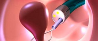
A tissue sample taken via biopsy is subjected to immunohistochemical analysis. This is a method of collecting biomaterial using a thin needle. It is injected into the patient’s body and the tumor tissue is drawn in using a syringe system. The sample is being prepared:
- Fixation with paraffin.
- Making slices.
- Distribution of sections across glasses.
- Treatment of samples with antibodies and dyes.
Before examination, the biomaterial is stored in formaldehyde. This allows a single sample to be used for a long time without injuring the patient. A biopsy can be painful if the tumors are located in hard-to-reach places. If possible, malignant tumors in the brain are removed promptly, and the tumor itself is analyzed. It is important to preserve the material, since tumors can be up to 1 cm in size. Based on the test results, a treatment program is built. However, in this case, immunohistochemistry has an advantage.
Working with biopsy material
The essence of the study
An immunohistochemical test consists of examining tumor tissue cells, which is carried out using special reagents. These reagents contain antibodies - specific proteins labeled with certain substances that can be detected (for example, fluorescent dyes, enzymes, colloidal gold and others).
Antibodies have a specific spatial structure that allows them to bind to the antigen - the substance of interest in tumor cells. This chemical reaction is called “antigen-antibody” and indicates the presence of tumor-specific substances in the sample taken.
In particular, immunohistochemical studies may look for certain hormone receptors (for example, estrogen, progesterone and others), cancer markers, markers of increased cancer cell growth and other antigens.
In clinical practice, the most commonly used method is indirect immunohistochemistry, when antigens obtained from patient tissue are associated with primary antibodies. This creates an “antigen-antibody” complex, to which secondary antibodies are attached. Such a complex can be stained with special dyes, and specialists assess the status of the tumor based on the intensity of the staining.
Positive and negative sides
Advantages of the method:
- A minimum of biomaterial and reagents are required;
- Detects cancer at an early stage;
- Quick results;
- Accuracy and reliability.
An additional advantage is the possibility of three-dimensional modeling. The antibody response is visible after three hours. The final conclusion is given in 7-10 days. After this time, the type of cancer and stage of the disease, as well as its primary focus, will be accurately determined. Changes in the color of the markers usually do not cause discrepancies when making a diagnosis. A transcript of the results is written on the final form.
Reliability depends on the method of staining the sample with reagents. This is done automatically and manually. With the manual method, the human factor and laboratory technician errors cannot be excluded. The attending physician will tell you which laboratory to contact. The patient can independently inquire about how the analysis is carried out in a particular institution. To ensure the reliability of the result, it can be repeated or in two laboratories.
The resulting error is the fault of the patient. Before the biopsy, you should avoid hormonal drugs and other medications, exercise, and adhere to a diet. External factors influence results.
Application of the method in diagnostics
Indications for analysis:
- Pathologies of the reproductive system, infertility;
- Active tumor growth of any location.
There are no contraindications to the analysis. Sometimes it is impossible to obtain biomaterial for research due to the location of the tumor.
Endometrial immunohistochemistry is an important study for finding the causes of infertility. Its goal is to find antisperm cells that prevent fertilization. The cause of infertility is the insensitivity of the endometrium to hormones during ovulation. At the same time, hormonal levels are normal, there are no antisperm bodies. Immunohistochemical diagnostics reveals that the problem is in the endometrial receptors.
Thymoma is difficult to diagnose. Their division into malignant and benign is arbitrary. It all depends on the specific case. The status of the tumor of the thymus gland, thymus is shown by testing for antibodies.
IHC in rectal cancer is used to determine the epithelial or endocrine nature of the tumor, hereditary factor and prognosis. The technique determines whether the disease is primary and metastases.
The interpretation of the results is based on the degree of malignancy. The higher it is, the less favorable the prognosis. Pathology – 85% of the marker content means the last stage of the disease with metastases. Less than 10% is the norm, a successful outcome of treatment.
Other marker methods
An oncological diagnosis is still rarely made based on the results of a single study.
Histological
The second informative analysis is histology. The method differs from immunohistochemistry in that it gives an idea only of the cellular structure. Histologically, several different structures are identified in one material, for example, adenocarcinoma. A malignant formation is formed from glandular tissue that secretes fluid. Based on the type of fluid, serous and mucosecretory are distinguished. Adenocarcinoma cells are located follicularly or papillarily. The tumor can be solid or with inclusions of cysts, and is already classified as cystadenocarcinoma. These characteristics are learned from the results of histological examination and treatment is adjusted to the specific type of tumor.
The technology is the same as for immunohistochemistry. The material is biopsied, covered in paraffin, and the sample is cut into thin slices. The finished preparations are also stained, after which the pathologist examines them under a microscope.
The disadvantages of histological examination are the long waiting times for the result of a planned analysis, 10-14 days. Urgent analysis is done in 30 minutes.
Colored preparation for study
MGM method
Another analysis based on the reaction to markers is molecular genetic. Helps to identify abnormalities in the DNA of a malignant tumor that affect its metastatic and invasive activity, as well as the presence of a hereditary predisposition or the sporadic, spontaneous nature of the formation.
Histology and immunohistochemistry are the two main methods for making a diagnosis of cancer, confirming each other. Differences in results are rare because immunohistochemistry involves examination of cellular structure.
Often, after histological examination of the lymph node, the patient is additionally referred for immunohistochemistry. The material is examined under a microscope by an expert who may miss something. Or the structure is difficult to identify. The IHC method is a help in a controversial situation.