In a normal state, a woman should not have discharge from the mammary glands, except during pregnancy and lactation during breastfeeding. Therefore, if a transparent yellowish, brown, or green liquid is released from the mammary glands, or if painful sensations appear in the breasts, then it is necessary to urgently consult a gynecologist or mammologist - such manifestations may indicate a serious pathology or even cancer. In some isolated cases, there may be minor discharge that is not accompanied by pain or any other discomfort. Such discharge can be considered quite normal, but you still need to get it checked.
In developing countries, the diagnosis of breast carcinoma is still made by fine needle aspiration cytology. The prognostic information from cytomorphology conveyed to the clinician depends on the way the cytopathologist formats the report. This review focuses on cytomorphological features and various gradient systems with their strengths, limitations and practical applicability. Key words: breast, carcinoma, cytological grading, fine needle aspiration.
Cytology for breast cancer
Accurate diagnosis of breast cancer is made 99% of the time through a combination of clinical examination, mammography and a simple, non-invasive, cost-effective outpatient procedure, fine-needle aspiration cytology. On the contrary, in developing countries like India, even today, core needle biopsy is still hardly practiced in most medical centers. Prognostic markers important for treatment decisions should be conveyed to the surgeon, as recognition of disease aggressiveness is key to effective medical management of breast cancer and avoidance of unnecessary morbidity. This review discusses the various classification systems proposed by different authors over the years for cytology of breast carcinoma.
Usually, smears of mammary gland discharge are taken for cytological examination. Using this analysis, the cellular composition of the liquid is determined and the characteristics of the taken biomaterial are assessed.
Indications, technique, histology interpretation
Histology of the mammary gland is prescribed to determine the chemical composition of the cells of the resulting tissues, as well as their sensitivity to sex hormones. Studying the rate of division helps to understand how quickly the pathological process spreads. Indications for histological examination and biopsy:
- detection of lumps and nodes in the mammary gland;
- structural changes in the nipples;
- bloody and other discharge;
- redness, peeling, formation of ulcers or lemon peel in areas of the mammary gland;
- detection of suspicious areas on ultrasound or mammogram;
- spots on an x-ray.
In most cases, diagnostic appointments do not confirm the presence of dangerous cancer cells. However, histology is necessary for everyone who is at risk:
- the presence of intraductal papillomas, cysts, fibroadenosis;
- diffuse tissue hyperplasia;
- abnormal growth of compaction;
- necrosis of tissues, formations inside the structure;
- stage 1-3 cancer and metastases;
- breast deformation.
Many patients are worried when they receive a referral for histological examination. However, this is only excessive stress, because in 85% of cases where cancer is not detected, the diagnosis indicates the development of completely different diseases.
During the examination of the obtained material, a high-precision microscope is used, which can show: the stage of oncological pathology, the spread of the formation, the morphological nature, as well as the hormonal characteristics of the tumor.
Based on the characteristics of cellular structures, diagnosticians can distinguish the forms and types of cancer tumors:
- tubular cancer with tube-shaped cells;
- medullary cancer when cells coincide with brain tissue;
- mucous cancer in the presence of mucin in the cells.
Histological examination also helps to distinguish the oncological process from benign pathologies, in which structureless masses can be observed in the punctate of the mammary gland.
To obtain a histological sample, 2 methods of intervention are used - using a needle or excision. The operation is performed on an outpatient basis; no preparation is required. Only avoid aspirin, if possible, 1-2 weeks before the procedure. A biopsy is performed as follows:
- The woman is placed on the couch on her side or back.
- Local anesthesia is given.
- An ultrasound device or x-ray is used to mark the site of excision or needle insertion.
- An incision or puncture is made and material is taken.
Then stop the bleeding, apply ice and a tight bandage. The procedure takes a maximum of 60 minutes, after which the patient is discharged home.
After taking the biomaterial, the study begins:
- The composition is placed in a formaldehyde solution and sent to the laboratory.
- The tissues are dehydrated to increase density, then embedded in paraffin.
- Using a microtome, the tissue is cut into plates and placed on slides for diagnostics.
- Using enzymes, dyes and antibodies, the specialist studies the nature of the cells and the boundaries of the tumor.
After the histological examination, the laboratory specialist draws up a conclusion and sends it to the patient’s attending physician.
The laboratory usually studies the sample within 5-10 days, after which the result is immediately sent to the doctor. If the institution where the patient underwent the biopsy has its own laboratory, the result will be ready in 2-3 days.
An expert opinion is issued in writing. It contains a description of the anomalies or lack thereof. Various digital data and indicators of markers are also given. Sometimes names in Latin and medical terms are prescribed to denote a particular process.
It is difficult for a person without the appropriate education to understand the terminology presented, so the patient should definitely consult a doctor. Interpretation of the results is tied to the definition of malignancy - its degree and nature. The degree of differentiation of the neoplasm is also assessed. These factors make it possible to accurately determine the following parameters and levels of malignancy in the mammary gland:
- GIV – indicator of the maximum stage of the malignant process;
- G – minimum degree;
- GX – confirmation of the benign nature of the tumor, which is usually expressed as a fibroadenoma.
Additionally, the analyzes will contain the following data characterizing certain markers:
- ER+ and PR+ - positive indicators for progesterone and estrogen, which indicate the hormonal dependence of tumors;
- ER- and PR- - a negative result helps to find out the likelihood of relapse and select hormonal therapy.
If HER2 positivity is detected, it is a positive, active cancer that requires surgery and chemotherapy. HER2 negative tumors are less aggressive and respond better to treatment. Also, when studying the test results, the specialist identifies Ki-67, which, if the reaction is positive, indicates low sensitivity to hormones, which is a direct indication for chemotherapy.
Histological examination of the mammary gland is an accurate way to diagnose existing neoplasms in order to establish their type, mode of development and dependence on hormones and other factors. Histology is necessary to prescribe effective treatment.
Pathologies of glandular tissue
Various pathological conditions in the glandular tissue of the breast, as a rule, develop with the following diseases:
- neoplasms of the ovaries or uterus;
- endometriosis;
- polycystic ovary syndrome;
- uterine fibroids;
- disturbances in the functioning of the endocrine system.
The pathological condition is accompanied by active tissue proliferation and hormonal imbalance. The development of the disease can be provoked by a number of internal and external negative factors, as well as hereditary (genetic) predisposition. The disease can begin to develop after mechanical impact on the sternum, stress, nervous exhaustion and prolonged use of hormonal drugs, especially if they are not suitable for the person. There have been cases where the disease actively progressed in people who interacted with dangerous chemicals.
Clinical picture of the development of hyperplasia:
- secretion of secretions of different colors, smells and consistency from the nipple;
- recurrent or constant chest pain;
- swelling of soft tissues;
- upon palpation, nodes are clearly visible.
The diffuse form occurs without a pronounced clinical picture, so it is extremely difficult to differentiate the disease. In most cases, the diagnosis is made during a routine examination by a mammologist.
In medicine, hyperplasia of glandular tissues is divided into several types.
| View | Peculiarities |
| Dishormonal | Develops against the background of hormonal imbalance. |
| Atypical | The condition is medically classified as precancerous. It is characterized by active proliferation of glandular, adipose and epithelial tissues. |
| Epithelial | Characterized by damage to the epithelial layer. |
| Stromal | Accompanied by the formation of a benign fibromuscular tumor. |
| Nodal | The progression of the pathology is accompanied by the formation of nodes. |
| Ductal | Active tissue growth occurs in the milk ducts. |
| Diffuse | Characterized by the formation of multiple granular tumors. |
| Cystic | During the examination, single and multiple cysts are detected. |
| Fibrous | Dense neoplasms that are fused with the surrounding tissues. |
The pathology, despite the vague clinical picture, requires treatment.
This pathological condition is accompanied by severe pain in the chest. The woman is worried about heaviness and discomfort. Sometimes the pain radiates to the armpit. Spontaneous discharge mixed with pus may be observed from the nipple.
In medicine, there are several forms of the disease:
- fibrous-adenomatous – the condition is characterized by the proliferation of organic structures;
- glandular-cystic - cysts form in the layers of soft tissue;
- glandular-fibrous – benign formations are formed.
Each form has its own characteristics of course, development and treatment.
When the first symptoms of pathology appear, you must contact a medical facility. Glandular mastopathy, as a rule, does not affect lymph nodes.
Indications, technique, cytology interpretation
Breast cytology is prescribed in cases where the first suspicion of pathological processes in the breast appears. This diagnostic method gives a reliable result in 97% of cases; the indications for it are:
- suspected infection, inflammation or cancer;
- confirmation of oncology during resection;
- evaluation of treatment effectiveness results;
- prevention;
- monitoring the condition after treatment or when the patient enters a risk group;
- undiagnosed infertility;
- pain and discomfort in the mammary glands.
It is recommended to take cytology only after preparation and only on days 6-14 of the cycle. If you break this rule, the procedure may be painful. During menopause, cytology can be performed any day.
A week before cytological examination of breast punctate, it is necessary to stop taking aspirin and anticoagulants. On the day of the procedure, you should not apply perfume or deodorant to the chest area. There is no need to remove your bra. However, before diagnosis, you need to wash your breasts. If a woman is very worried, you can take a sedative. Receive material in the following ways:
- Aspiration biopsy. It is prescribed for small tumors - up to 1.5 cm in diameter and only if they are located in the upper layers of the mammary gland.
- Core biopsy. Recommended if the tumor lies deep in the structures. To obtain the material, use a gun with a thick needle.
The duration of the procedure is from 5 to 10 seconds, no longer is needed.
The study in the laboratory occurs in several stages, they last up to 2-3 days. In urgent cases, cytology is performed within 1 hour. After receiving written data, the patient must consult a doctor.
- Incomplete data. If cytology is inaccurate, additional diagnostics may be required.
- Norm. The data will indicate that no deviations or anomalies were identified.
- The presence of benign cells. The analysis will show a large amount of connective tissue and structureless substances in the mammary gland with morphological abnormalities. Macrophages with red blood cells will also be detected.
- Non-cancerous cells. The sample contained non-cancerous cells, but abnormal ones: mastitis, cyst, inflammation.
- Atypical and malignant cells. The formation of a cancerous tumor, additional data about the location of the lesion, its boundaries, shape, stage, and structural features will also be indicated.
If a negative diagnosis is obtained by cytology, additional diagnostics may be required. Cuboidal epithelial cells of the mammary gland may be detected, which also require additional examination.
Cytological and histological examination of the mammary gland are similar procedures that differ in the technique of execution, although not significantly, as well as in the method for determining the results. Most often, the first method is used with minimal suspicion and the absence of significant symptoms of a tumor, as well as for monitoring and prevention after treatment. Histology is used for detected tumors. The method and method of diagnosis can only be determined by a doctor.
- How does breast cancer occur?
- Types of Breast Cancer
- Causes and risk factors
- Symptoms of breast cancer
- Self-diagnosis of breast cancer
- Diagnostics
- Stages of breast cancer
- Breast cancer treatment
- Prognosis for breast cancer
Indications for the study
Cytological examination of smears of discharge (discharge) from the mammary glands is a very informative and reliable analysis - the result guarantees 90-97 percent accuracy. Indications for taking smears for cytological examination are the following circumstances:
The way the cytopathologist formats the report determines whether accurate and complete information based on cytomorphology is conveyed to the clinician or not. The cytological grading mentioned in the cytology report actually adds to the objectivity, reproducibility and authenticity of a particular report. In addition, importance has been given to a cytological grading system that will closely correspond to the grading system used in the histological material, but the most reliable method of cytological grading remains to be determined.
- Neoplasms in breast tissue can be both benign and malignant processes.
- Any discharge from the breast that is not associated with lactation or pregnancy.
- In case of a change in the color of the skin of the mammary glands, the appearance of flaky areas, or a violation of the integrity of the skin.
- For dyshormonal pathologies such as mastopathy or mastitis.
- Chronic diseases of the female reproductive system.
- Traumatic injuries to the mammary glands: bruises, wounds.
- Any inflammatory processes in the mammary gland.
Smears of discharge (discharge) from the mammary glands for cytological examination are taken when visiting a gynecologist, mammologist or oncologist.
The different classification systems used for breast cancer cytology are compared. The study focuses on the cytomorphological features and scoring criteria reviewed by various authors for reporting breast carcinoma. Various evaluation methods were discussed for their strengths, short-circuits and practical applicability.
The present study includes information collected from each article regarding the architectural and cytological features examined in breast smears, the method and objective scoring criteria. The classification criteria and cytomorphological features considered by various authors are tabulated and discussed for their usefulness and limitations. All classification methods were developed for the most common type of breast cancer, that is, infiltrating ductal carcinoma.
Every woman should know that smear examination is an important part in diagnosing any pathology of the mammary glands. With the help of cytology, modern medicine quickly and accurately detects cancer in the early stages, and this gives a great chance of successful treatment.
Discharge from the mammary glands may be constant or appear periodically. Depending on the disease, the color of the discharged fluid may vary. For example, green color is characteristic of mastopathy, brown indicates slight bleeding in the milk tubules, purulent discharge is a signal that there is an inflammatory process in the breast.
Most researchers have used Pap lesions for classification purposes. The number of cases varied from 20 to 281 in these studies, while category 2 cancers included the majority of cases among the study groups. Nuclear features were invariably included in the scoring scheme of all investigators.
Various assessment methods. Tumor cytology assessment is quick, easy to perform, and correlates well with core. Cytological classification of breast carcinoma - Possible proposal? Tumor cell continuity as a prognostic factor in aspiration smears of breast carcinoma.
How does breast cancer occur?
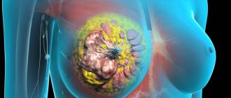
Breast cancer develops in the same way as any other malignant tumor in the body. One or more cells of glandular tissue, as a result of a mutation that has occurred in them, begin to divide abnormally quickly. They form a tumor that can grow into neighboring tissues and create secondary tumor foci - metastases.
Mutations that lead to breast cancer can be hereditary or acquired.
Common inherited genetic causes of breast cancer are mutations in the BRCA1 and BRCA2 genes. BRCA1 mutation carriers have a 55–65% risk of developing breast cancer, and BRCA2 carriers have a 45% risk of developing breast cancer. Such genetic defects are inherited from parents to children and cause breast cancer in approximately 15% of cases.
Much more often, a tumor occurs due to acquired mutations: they arise in the cells of the mammary gland and are not inherited. For example, in 20% of cases, the number of copies of the gene encoding HER2, a receptor protein that is located on the surface of cells and stimulates their reproduction, is increased.
Main criteria
The main criteria for this study are cells, nuclei and nucleoli.
Signs of disturbances in the nucleus in cancer:
- The size of the core is noticeably increased.
- The kernel is tuberous.
- Lack of an even contour.
- Polymorphism.
- Uneven chromatin pattern.
Signs of cell abnormalities in cancer:
- Increase in cell size.
- Changed form.
- Change in the proportions of the cell's cytoplasm and nucleus.
- Discrepancy between the parameters of cytoplasmic maturity and the parameters of nuclear maturity.
Signs of disturbances in the nucleolus:
- The size of the nucleus is significantly increased, or it has an irregular shape.
- Increased number of nucleoli compared to a healthy cell.
Criteria for a malignant disease are often detected, but there are cells in which this criterion is not present or is weakly expressed. In such cases, a cytologist studies changes in the nature of cellular relationships. The analysis of the result and the conclusion are made on the totality of all the signs.
Types of Breast Cancer
Malignant breast tumors are divided into two types: ductal and glandular. Ductal breast cancer is more common. It can be intraepithelial (in situ) and invasive. Intracellular ductal breast cancer has a more favorable prognosis, it rarely metastasizes and is curable in 98% of cases. The invasive version of the tumor is prone to uncontrolled growth and generalization of the process.
Glandular breast cancer can be lobular (invasive lobular carcinoma) or grow from other cells of the glandular tissue. Lobular cancer is often characterized by multicentric growth. The rate of increase in size and timing of metastasis of forms of nodular breast cancer depend on the degree of tumor differentiation.
Forms of breast cancer
Approximately 70% of breast cancer cases are classified as invasive ductal. 10% of cases are cancer of the combined ductal-lobular structure. About 10% are rare forms: signet ring cell, secretory, lipid-secreting, colloid, squamous cell, Paget's cancer, apocrine.
Favorable prognoses are observed for tubular, juvenile, medullary, and adenoid cystic forms. The infiltrative lobular form of breast cancer reduces the favorable course of the disease. In lipid-secreting, infiltrative ductal, signet ring cell forms of the disease, researchers give a poor prognosis.
Causes and risk factors
Unfortunately, scientists do not yet have complete information about the causes of breast cancer. There is a list of risk factors that influence the likelihood of a tumor, but in some people the disease is diagnosed in the absence of these factors, while others remain healthy in the presence of many of them at once. However, scientists still associate the development of breast cancer with certain circumstances that most often precede its appearance. These include:
- Age. The majority of breast cancer cases occur in women aged 55 years and older.
- Heredity. If breast cancer is diagnosed in one of your close relatives, the risk doubles.
- History of breast cancer.
- Increased breast tissue density as determined by mammography.
- Some benign neoplasms in the mammary gland.
- Early onset of menstruation - before 12 years of age.
- Late menopause - after 55 years.
- Lack of children or late (after 35 years) first birth.
- Exposure to radiation, such as from radiation therapy given to treat another type of cancer.
- Smoking and alcohol abuse. If a woman consumes 28–42 g of ethyl alcohol daily, her risks increase by 20%.
- Excess weight and low physical activity.
- Use of hormonal drugs: oral contraceptives, hormone replacement therapy in postmenopause.
- Breast injuries.
- Diabetes.
- Work on a schedule with night shifts.
Macrophages in the thyroid gland:
Macrophages and neutrophils in the thyroid gland are also found in large numbers in diseases. A fairly common pathology of this organ is a cyst. After making a puncture (puncture) of the cyst, specialists obtain fluid. There are few cells in it, among them macrophages with hemosiderin predominate - they are also called siderophages.
Also, macrophages in the thyroid gland can occur in other diseases, for example, during an inflammatory process. However, in autoimmune thyroiditis, which is the main inflammatory disease of this organ, there are few such phagocytes in the biopsy specimen - lymphocytes predominate in the gland, and other immune cells are less common.
So, the presence of a large number of macrophages in the organs described above is a sign of a pathological process. Although, in order to make an accurate diagnosis, one must pay attention not only to how many macrophages are in it, but also to the presence and quantity of other components. Only by assessing the occurrence and correlation of all identified components can we draw a conclusion about what is happening to a person and what to do to restore his health.
By the way, when it comes to recovery, it is important not only to directly influence the cause of the disease (oncological, inflammatory, dishormonal process), but also to pay attention to supporting the immune system, of which the notorious macrophages are part. Taking the drug Transfer Factor allows you to normalize the functioning of the immune system and help macrophages in carrying out their good work - fighting pathogens and other harmful factors.
Regular therapy with this remedy can help you recover from most diseases of the mammary and thyroid glands, and when taken prophylactically, prevent their occurrence. With stable and coordinated work of all parts of the immune system, which is achieved thanks to Transfer Factor, the risk of developing autoimmune, inflammatory and even oncological processes in these organs is greatly reduced.
Mastopathy and breast cancer are fairly common diseases.
To identify the presence of pathological processes in the mammary glands, a number of examinations are carried out, including cytological analysis. Sometimes the result shows the presence of monocytes. This frightens and worries many women.
It is important to understand what macrophages in the mammary glands mean.
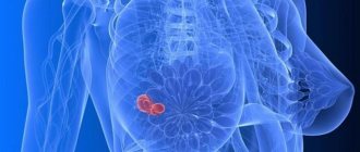
Symptoms of breast cancer
In the early stages, breast cancer usually does not manifest itself clinically. Most often, the tumor is discovered by the patients themselves or is detected accidentally during preventive studies.
Patients usually complain of the presence of a palpable formation and discharge from the nipple. Pain is a rare symptom of breast cancer, but the pain syndrome can come to the fore at the stage of generalization of the process, especially when metastases spread to the bones.
Quite often, signs of breast cancer are detected, such as the appearance of asymmetry due to changes in the size of the affected gland. Reduction, upward displacement, deformation and wrinkling of the mammary gland can be observed in the scirrhosis (fibrous) form of the tumor. On the contrary, the breast on the affected side becomes enlarged due to the rapid growth of the formation or due to edema, which forms due to impaired lymph outflow.
When the tumor spreads into the subcutaneous tissue, skin changes may be observed. The following symptoms of breast cancer are identified:
Sometimes, when the tumor spreads to the surface of the skin, signs of breast cancer such as redness and ulceration may be observed. The presence of these symptoms indicates that the process is neglected.
Changes in the nipple can also be detected, but only in the later stages. In this case, the following symptoms of breast cancer occur:
- Forga's sign - on the affected side the nipple is higher than on the healthy side.
- Krause's sign - the nipple is thickened, the folds of the areola are noticeably pronounced.
Such a sign of breast cancer as pathological discharge is quite rare, but in some cases it may be the only symptom that is detected during examination. The discharge is often bloody in nature, serous and purulent are less common.
Special forms of breast cancer have also been identified, which manifest themselves with typical symptoms. These include:
- An edematous-infiltrative form, which is characterized by enlargement and swelling of the gland, marbled skin color, and severe hyperemia.
- Mastitis-like. This type of breast cancer is manifested by hardening of the affected breast and increased body temperature.
- An erysipelas-like form, in which lesions are detected on the skin (sometimes ulcerations appear) that externally resemble erysipelas.
- The armored form is characterized by the presence of multiple nodes, due to which the gland shrinks and deforms.
- Paget's cancer affects the nipple and areola. With this type, thickening of the nipple, changes in the skin in the form of redness and thickening, and the formation of crusts and scales are observed.
Sometimes people, wondering what signs can be used to recognize the presence of a breast tumor, mistakenly look for symptoms of sternum cancer. This name is incorrect, since the sternum is the central flat bone of the chest and, even with metastasis of a malignant breast tumor, is almost never affected.
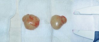
Breast biopsy: how is it performed and what does it show?
The issue of identifying and treating cancer is relevant today. There is no effective therapy yet that could help cure cancer at any stage.
However, early detection of the disease promotes rapid recovery. A breast biopsy can detect pathological cell disease at an early stage.
The procedure is carried out according to a special algorithm, showing not only cancer, but also other pathologies of the gland.
The site zheleza.com recommends that every woman, from the moment of puberty, be examined annually by a gynecologist and mammologist. The reproductive system and mammary glands are interconnected. Detection of the disease at the stage of absence of symptoms that would indicate it allows you to quickly prescribe therapy, which will be more effective than at a later date.
A biopsy is the insertion of an instrument into the body, through which a small part of the affected tissue is removed. The cells are placed on a glass slide, listened to, stained and examined for pathological changes. The operation can be performed under both visual and ultrasound supervision.
Mammography can detect the presence of cancer, but not completely. Some cells may not be recognized on film. However, a biopsy helps in identifying the disease 100%.
A biopsy is prescribed only after observation by a mammologist, who will first conduct an external examination and prescribe preliminary tests. Indications for a biopsy are predominantly darker or lighter areas than the surrounding tissue, as well as various lumps in the mammary gland. The doctor also explains how to prepare for the procedure.
A woman should contact a mammologist if:
- Papillary discharge.
- Dense internal formations.
- Peeling of the skin of the chest.
- The appearance of blemishes on the skin.
The doctor conducts an initial diagnosis and prescribes a biopsy if:
- The picture shows the seals.
- There are strange shadows on ultrasound or mammography images.
- Ulcers appeared, there was bloody discharge from the nipples, and the shape of the breasts changed.
With early diagnosis using biopsy, malignant tumors are detected in 20% of cases, and benign in 80% of cases.
Biostimulants, pregnancy or breastfeeding by a woman are contraindications for a biopsy. If a woman is allowed to undergo the procedure, then the doctor discusses with her how to prepare for it:
- 2-3 days before the biopsy, exclude drugs that especially affect blood clotting.
- Purchase a bra that the woman will wear after surgery and that will hold ice on the puncture site.
- Do not use cosmetics on the day of the procedure, so that the substances do not get into the open wound and give distorted results.
go to top
Fine needle biopsy is one of the most common today. This procedure is quite painless and local anesthesia is possible. Using a thin needle, the doctor penetrates the affected tissue and takes a liquid suspension. Next, the biomaterial is sent for cytological examination. If the formation is hard, then a thicker needle is used.
Aspiration biopsy sometimes does not give accurate results. A false negative result is considered if no cancer cells were found in the sampled tissue.
The fact is that a cancer tumor can be heterogeneous: it can consist of malignant and benign cells.
To more accurately establish the disease, the entire damaged area is surgically removed using a scalpel, and then the composition of the cells can be seen under a microscope from many sections.
A mammogram is performed before the biopsy, which allows you to rule out the procedure or not. An aspiration biopsy is most often performed rather than a surgical one.
To perform a biopsy with a thicker needle, a hollow instrument and mammography are taken. The specialist inserts a needle into the affected area while looking at the image.
Once the needle touches the affected area, a marker needle will be inserted and will remain there until the end of the operation. Biomaterial is taken and examined.
Core biopsy refers to the use of a thicker needle, which allows you to take an entire area of pathological tissue and examine it. Thus, biopsies are performed with different instruments with different diameters.
go to top
There are other methods of performing a biopsy. A stereotactic biopsy is performed with a single needle in several areas of the affected area. To accurately target the tissue, an ultrasound or digital mammography is used. This biopsy allows you to identify changes in tissue structures, determine the nature of the formations, and not damage calcium formations at the surgical sites.
Stereotactic biopsy is considered as accurate as surgical biopsy. Its advantage is that it leaves no scars or defects, unlike surgical intervention. Moreover, it is painless.
Vacuum biopsy is innovative because it allows both diagnosis and therapy to be carried out simultaneously. Material is taken from several areas in volumetric samples using ultrasound or x-ray. This method is considered better than trephine biopsy or puncture.
With a vacuum biopsy, the needle is inserted once and rotates around its axis, taking material into the hole using vacuum. This method is less traumatic and more informational, since it allows you to take material from various areas, causing the least amount of damage to the skin of the chest.
Vacuum biopsy can also be used as a therapeutic method to eliminate benign tumors. If a woman is diagnosed with a malignant tumor, then this procedure is not used instead of surgery.
go to top
Trephine biopsy, or trephine biopsy, allows you to determine the type and size of the tumor, the stage of cancer development. Here, a tubular needle is inserted directly into the tumor, from which fluid is removed down to the walls. By removing the tumor completely, the disease can be accurately diagnosed.
Trephine biopsy, like puncture biopsy, is performed before radiation treatment, when an accurate picture of what is happening is needed. You need to know the structure, size and other components of the tumor in order to cure it.
Fine needle biopsy is the easiest and most common. The woman is given local anesthesia, a needle is inserted under the skin and biomaterial is taken. However, this method has a disadvantage when the material is taken from only one area of the pathological formation.
go to top
Research results
The results of studies after a biopsy are histological and morphological, divided into the following types:
- Incomplete - when the picture remains incomplete due to insufficient material or due to an inaccurate sample.
- Normal - when the material provided comprehensive information to laboratory technicians, which does not require additional research.
- Benign – when the examination reveals the corresponding cancer cells.
- Malignant - cancer cells have a period and stage of development, there are clear boundaries, as well as symptoms of the disease.
go to top
Self-diagnosis of breast cancer
You should check your breasts yourself for the presence of nodules or any other changes once a month after menstruation. It is most convenient to carry out home diagnostics while taking a bath or while under the shower. You should tell your doctor about any changes that are discovered as soon as possible.
Procedure for performing breast self-examination:
- Undress from the waist up and stand in front of a mirror.
- Raise your hands up and place them behind your head. Examine your breasts carefully. Turn right, left side.
- Feel the mammary glands while standing with your index, middle and ring fingers folded together. Start at the upper outer part of the chest and move clockwise.
- Pinch the nipple with two fingers. Check to see if anything stands out.
- Feel the mammary glands again - now in a lying position.
70% of breast cancer cases are self-diagnosed by patients through breast self-examination.
Functioning of macrophages
Macrophages (or mature monocytes) are a type of white blood cell.
Together with monoblasts and promocytes, they form a system of mononuclear phagocytes. These are immune cells. They are vital for stimulating the work of the body's nonspecific defense mechanisms. Their activation leads to the absorption of intracellular pathogens: Listeria, Mycobacterium tuberculosis, Toxoplasma, Leishmania, Streptococcus pneumoniae, etc.
The destruction of such pathogens depends on the production of nitric oxide and hydrogen peroxide. Macrophages are present in almost all tissues of the body, including the mammary glands. Their main role is to cleanse the body of damaged, cancerous and dead cells, elements, bacteria and other pathogenic bodies.
The process by which macrophages engulf and disarm unhealthy tissue is called phagocytosis. Macrophages are involved in maintaining adaptive and cellular immunity. They receive information about foreign antigens and transmit it to lymphocytes. Thanks to this, the immune system is able to better cope with the pathological process.
Macrophages also take part in the following important body functions:
- wound healing;
- homeostasis;
- cessation of immune reactions;
- production of hormonal substances.
Immune cells can be in a calm or activated state, the latter state provokes:
- complement components (complex proteins that are constantly present in the blood);
- bacterial products (LPS);
- adhesion to different surfaces;
- cytokines.
Activated macrophages differ from passive ones in a number of morphological characteristics, for example:
- adhesion ability increases;
- increased cytotoxic activity;
- big size;
- degradation of trapped particles;
- formation of products of partial reduction of oxygen;
- increased secretion of monokines, lysosomal enzymes;
- active expression of various receptors (to cytokines IL-1, 2, 6, transferrin, TNF).
Diagnostics
Diagnosing breast cancer begins with a conversation. At this stage, it is important for the doctor to evaluate the woman’s complaints and find out whether cases of breast cancer have occurred in her family, and if so, how often. This helps to suspect a hereditary form of cancer associated with mutations in the BRCA1, BRCA2, NBS1, CHECK, TP53 genes.
Next, the doctor examines, palpates the mammary glands, checks to see if there are any nodes or lumps in them, or if the lymph nodes in the axillary, supraclavicular and subclavian areas are enlarged.
After the examination, the doctor may refer the woman for a mammogram, an X-ray of the breast. Indications for this study are: lumps in the mammary gland, changes in the skin, bleeding from the nipple, as well as any other symptoms that may indicate a malignant tumor. Ultrasound examination is also prescribed to diagnose breast cancer. Mammography and ultrasound are complementary methods, each of them has its own advantages:
Mammography
Ultrasound of the mammary glands
Allows you to detect pathological changes 1.5–2 years before the onset of symptoms.
If there is bloody discharge from the nipple, ductography can be performed - X-ray with contrast of the milk ducts. This helps to obtain additional useful information.
High sensitivity - accurate diagnosis of up to 90% of cancer cases.
Ability to detect microcalcifications up to 0.5 mm.
Safety - there is no effect on the body from x-rays.
Well suited for high density breast tissue in young women (up to 35–45 years).
Allows you to distinguish cysts (cavities with fluid) from solid tumors.
Allows you to assess the condition of regional lymph nodes.
Well suited for monitoring needle position during biopsy.
Magnetic resonance imaging is a highly informative method for diagnosing malignant breast tumors. It is used for lobular cancer, when mammography and ultrasound are uninformative, as well as for assessing the size and location of the tumor, which helps determine the tactics of surgical treatment. MRI can be used to screen women who carry abnormal genes associated with an increased risk of breast cancer and have a strong family history.
Doctor of the European Clinic Portnoy S.M. talks about the role of biopsy in diagnosing breast cancer:
In the laboratory, cytological and histological examinations are carried out, that is, the structure of individual cells and tissues is assessed. Molecular genetic studies are currently available: they help to identify mutations that caused malignant degeneration and to select optimal antitumor therapy.
A biopsy can determine whether a tumor is cancerous, as well as determine its type and stage. In addition, examination of the biopsy material provides an answer to the question of whether the tumor is hormone dependent, which also affects the treatment regimen.
Once cancer is diagnosed, it is important to determine its stage and understand how far it has spread in the body. The following studies are used for this:
Cytology objects
Objects of breast cytology are:
- Cyst contents.
- Discharge directly from the nipples.
- Scrapings from lymph nodes.
- Breast tumors.
- Lymph node punctures.
- Scrapings from areas of the nipple or breast skin that have undergone ulcerative and eroded processes.
This research method is carried out by an ultrasound specialist or attending physician. If the object is nipple discharge, it is removed by applying pressure on or around the nipple area. The tissue of the entire mammary gland is also affected. When conducting diagnostics, the first and also the last drops of discharge are taken for analysis.
National Surgical Adjunct Breast and Bowel. The project showed that administration of tamoxifen reduced the risk of invasive and non-invasive breast cancer. almost 50% in all age groups. In a subgroup of patients with ductal atropy. prophylactic tamoxifen reduces the incidence of breast cancer by 86%. 7 There are also established algorithms to guide the situation with women. with an increased risk of breast cancer. 8.
Despite the presence of these preventive measures. approaches, traditional methods for assessing the risk of breast cancer are not very large. useful for identifying patients who may benefit from them. Between 50% and 70% of women who develop breast cancer have no identifiable risk factors. except age, using modern standard risk assessment tools. Statistical risk assessment methods - for example, the Gale model, which uses a woman. personal and family medical history to assess risk - more useful. epidemiological tool than for identifying personal risk. 9 For example, the Gale model is known to underestimate risk in some patients. population and often overestimate the risk in others. 10 Because these traditional methods have proven inadequate, they continue. interest in developing readily available biomarkers that can identify. risk of breast cancer with an acceptable degree of individual prognosis. accuracy.
If the punctate is a liquid, sodium citrate is added to it, otherwise it may curdle. The test object is then placed in a centrifuge, and smears are made from the resulting sediment. Next, the result is deciphered.
Principles of Cytology:
- Analysis of the composition of cells in normal conditions and pathological changes in breast cancer.
- Analysis of a group rather than a single cell. The background of the drug is important.
- A cytological examination always ends with the formulation of a conclusion.
Stages of breast cancer
Staging for breast cancer is based on the generally accepted TNM system. The T in this abbreviation indicates the size of the primary tumor:
The letter N indicates the presence of metastases in regional lymph nodes. N0 - there are no foci in the lymph nodes. N1, N2 and N3 - damage to different numbers of lymph nodes.
The letter M indicates the presence of distant metastases. One of two numbers can be indicated next to it: M0 - no distant metastases, M1 - distant metastases present.
Depending on the values of T, N and M, there are five main stages of breast cancer (within some of them there are substages):
- Stage 0: cancer in situ.
- Stage I: tumor in the mammary gland up to 2 cm in diameter.
- Stage II: a tumor in the mammary gland with a diameter of up to 5 cm or more, there may be metastases in the axillary lymph nodes on the affected side.
- Stage III: a tumor in the mammary gland up to 5 cm or more, can grow into the chest wall or into the skin, there are foci in the regional lymph nodes.
- Stage IV: The tumor can be of any size, it does not matter whether regional lymph nodes are affected. If distant metastases are detected, stage IV cancer is always diagnosed.
Intraoperative cytology
The intraoperative method is considered the basis of the cytological method. It reveals the pathology of the malignant process, as well as the spread of metastases to the liver and lymph nodes. The reliability of this method is about 98%.
Cytological examination of the mammary gland studies the pathomorphosis of treatment during photodynamic, as well as chemoradiotherapy. This method allows you to determine the nature and extent of the malignant process in the mammary gland even before surgery. The cytological method predicts the course of the disease and identifies factors that have a significant impact on determining the correct treatment regimens.
The reliability of this study is increased through the use of the liquid cytological method, as well as the introduction of special laboratories, the use of completely new microscopic systems that allow decoding and morphological analysis of the dynamics during the life of cells.
Breast cancer treatment
The treatment strategy for breast cancer should be selected individually for each patient, taking into account factors such as tumor type, stage, and sensitivity of the tumor to hormonal therapy. The general condition of the patient is also taken into account. If the tumor is detected in the early stages and the correct patient management tactics are chosen, then the chance of completely curing breast cancer is very high.
Select an oncologist and make an appointment:
Plastic surgeon, oncologist-mammologist, Doctor of Medical Sciences
Goals and objectives of cytology
The main goals when diagnosing the mammary gland using a cytological method are:
- Conclusion of specialists before the start of treatment.
- Fast diagnostics.
- Monitoring the degree of therapeutic effectiveness.
- Monitoring the course of the disease.
The diagnostic results using this method are compared with the results obtained in histology.
Diagnosis of breast cancer using cytology is considered one of the most accurate (90-96%). A small percentage of cases of such research are unsuccessful due to the unsuitability of the material for analysis.
Indications and contraindications of the method
Breast cytology is based on the study of the characteristics of the cellular material of tissues. This method is often used in a complex of diagnostic measures along with mammography, ultrasound, and MRI.
Indications for cytology:
- Suspicion of a tumor or other neoplasms.
- Nipple discharge.
- Presence of pain in the chest.
- Chest injury or bruise.
- Visible changes in the skin of the breast.
The method also has contraindications:
- Pregnancy
- Lactation period
- Increased body temperature
- Recent operations
- Poor blood clotting
- Infectious diseases in the acute stage
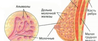
What is glandular tissue of the mammary gland, its functions
The mammary gland is divided into small lobules due to connecting layers, which diverge from the skin in different directions. The cavity located between the lobules is filled with fatty tissue. It plays the role of a kind of shock-absorbing pillow on which the gland itself is located.
The glandular tissue itself and its structure consists of a large number of small glands. They are located directly in the lobules of the mammary gland. Each section has tubes, the ends of which are slightly expanded. These small expansions are called alveoli.
Throughout a person’s life, the mammary glands undergo constant changes, for example, during puberty they actively grow. This condition is accompanied by the production of certain substances that stimulate the development and functioning of the breast. During menstruation, pregnancy, lactation and hormonal changes, a woman's breasts increase in volume and become more sensitive.
Every month during menstruation and after the end of regulation, the breasts undergo changes. With the beginning of the second phase of the menstrual cycle, the body begins to actively produce progesterone, which stimulates the growth of alveoli. At the end of the cycle, everything returns to its original state.
With the onset of pregnancy, the continued production of progesterone causes the alveoli to mature. In the last stages of pregnancy, the body begins to produce prolactin, which affects the state of organic structures. A woman’s body is intensively preparing for breastfeeding. With the birth of a baby, the hormone oxytocin begins to be produced, which regulates milk production. The functioning of the mammary glands is influenced by the work of the thyroid gland, adrenal glands and pituitary gland. In case of hormonal imbalances, it is necessary to normalize the balance with the help of medications.
During menopause and postmenopause, various transformations also occur in the breasts, the risk of developing various diseases increases, and the production of certain hormones in the body stops.
Cytological diagnostic methods
There are different ways to carry out diagnostics. The choice of method depends on the preliminary diagnosis and condition of the patient.
The material for research is cells, and they can be obtained in the following ways:
- Tissue scraping
- Nipple discharge
- Breast puncture
- Fabric prints
Methods of cytological examination:
- Puncture using fine-needle aspiration biopsy (FNA) is considered the simplest and least painful method. It is carried out in conjunction with ultrasound and mammography, which allows you to monitor the position of the needle as accurately as possible. During the study, anesthesia is not required, so it is used on an outpatient basis. The neoplasm is located by palpation. The doctor carefully treats this area with antiseptics and inserts a needle there. Using a syringe with a thin needle, take the required volume of material. Then the needle is removed, the puncture site is also treated with antiseptic agents and a bandage is applied. The resulting material from the syringe is placed on a glass slide or in a test tube with a special solution. If liquid oozes from the puncture site, it is collected in a test tube. Then, the doctor palpates the gland for residual discharge. The procedure is recommended to be done from 6 to 14 days of the menstrual cycle.
- Core needle biopsy of the breast - used mainly when a significant amount of material is needed for research, for example, if a cyst is suspected. This type of diagnosis involves the use of local anesthesia.
- Stereotactic breast biopsy - this method is carried out in combination with a mammography machine and is used in cases of deep-lying tumors.
- Trephine biopsy of the breast is a more serious and expensive method, performed under local or general anesthesia. It involves collecting a column of tumor tissue, which is taken with a thick, pointed needle. It produces more informative and accurate results, but is more traumatic and is not prescribed as often, but only in controversial cases.
- Cytology of discharge from the mammary gland. A healthy woman should not have nipple discharge unless it is associated with pregnancy or breastfeeding. Discharge from the mammary glands indicates the development of the disease. Cytology analysis shows the nature and cause of the disease. In this case, a smear or imprint of the secretions from both glands is taken. The required amount of exudate is placed on a glass slide. To keep the smear from drying out, special solutions are used.
- Liquid cytology of the mammary gland - this diagnostic method began to be used relatively recently; the punctate is placed in a special medium and processed in a centrifuge at 1000 rpm for 5 minutes. The material prepared in this way has a single-layer structure. It is evenly distributed over the surface of the slide, which facilitates examination and reduces time. This allows you to avoid the use of expensive serums, which significantly reduces the cost of the procedure. In addition to liquid cytology, there are options for fixing the material with special mixtures (Nikiforov’s mixture, acetone) or staining the materials (Papanicolaou, Romanovsky-Giemsa, Leishman, Pappenheim).
Preparing for the study
The procedure for taking smears of discharge (discharge) from the mammary glands does not require any special preparation from the woman. There are just a few rules to follow before research:
- a week before the procedure, stop taking antibiotics and antifungal drugs;
- if the patient is taking hormonal medications, the doctor must be informed about this;
- do not use deodorants or other aromatic products directly on the day of taking smears;
- Before going to the doctor, thoroughly wash your mammary glands.
Interpretation of the results of cytological examination
An accurate diagnosis depends on several conditions. Firstly, the quality of the procedure performed, secondly, the amount of material taken for the study and, finally, the correct interpretation of the test results.
Deciphering the results of breast cytology is an important final step in the process of making an accurate diagnosis and prescribing effective treatment.
The most common variants of cytological examination results:
- Normal - the material taken for analysis does not contain atypical cells, malignant formations, or inflammation in the tissues. The patient's health is not in danger.
- Incomplete results - yes, this also happens when the amount of material taken is insufficient or when controversial issues arise, in which case a repeat examination is ordered or another type of diagnosis is selected.
- Presence of cancer cells - the decoding must contain the abbreviation ASC (atypical squamous cells) and AGS (atypical glandular cells), structure, stages and localization of the tumor, degree of dysplasia.
- The accumulation of benign cells excludes the presence of an oncological neoplasm in the patient, but still indicates the development of pathology in the mammary gland.
Types of benign neoplasms:
- Intraductal papilloma, in which bloody discharge from the nipple, a small number of atypical cells, and rapid proliferation of cuboidal epithelial cells are observed.
- Galactorrhea - this pathology is characterized by such indicators as an increased level of leukocytes and erythrocytes, the presence of epithelial scales, and yellowish or greenish discharge.
- Fibrocystic mastopathy - in this case, the presence of xanthoma cells and foamy macrophages is possible.
- Fibroadenoma - a small amount of structureless substance and anucleate squamous epithelial cells may be detected.
The main advantages of cytological examination of breast punctate are:
- The method is absolutely safe.
- The procedure does not take much time.
- You don’t have to wait long for test results; on average, they are ready within three days.
- There will be no scars left on the body, and the hematoma formed at the puncture site will disappear in a few days.
- Low cost of the procedure.
- The accuracy of this diagnostic method is quite high and amounts to 90-95%.
Interpretation of the results obtained
The study of discharge (discharge) from the mammary glands plays the most important role in making a diagnosis and prescribing a further course of treatment. This technique makes it possible to determine the nature of pathological changes and the cellular composition of the secreted fluid. Interpretation of cytology results gives the doctor an idea of what measures to take, allows you to draw up the correct treatment plan and, if necessary, prescribe additional examination. A woman, having received the results of the study, should contact her doctor with them and not try to decipher them on her own. Only a specialist can understand what the analysis data say and determine what pathology we are talking about - an inflammatory process, infection, hormonal imbalance or cancer.
The need for an effective risk screen
Detection of atypia may alert the attending physician to further observation and, in some cases.
Preventive measures may be indicated. Emergence of new acquisition technologies. The past decade has made soil sampling more accessible to medical professionals. and more acceptable to patients. Rationale for breast health screening. using cytology as well as a review of collection methods. and guidelines for the interpretation and reporting of such samples. Although progress has been made in identifying and. treatment of breast cancer, the disease remains, except for skin cancer. the most common cancer for women in the United States. From all of the above, we can conclude that the appearance of discharge from the mammary glands in a woman that is not associated with pregnancy or lactation is a reason for immediate contact with a specialist (gynecologist or mammologist). Also, a woman should be prepared for the fact that, depending on the results of the smears taken of the discharge (discharge) from the mammary glands, she will be prescribed additional diagnostic procedures. This may include an ultrasound, mammography and blood test for hormones.
Discharge from the mammary gland
Despite the high total number of deaths.
The mortality rate from breast cancer has been declining in recent years. Improved detection methods are credited with much of the decline in mortality rates because they help find tumors earlier. this can be felt during a manual breast examination by a doctor or woman. herself. Early detection means the tumor may be smaller or larger. treatable, with fewer consequences. But mammograms are not very effective. Women with dense breasts, and they are not very useful if the goal. prevention of breast cancer. In addition, do not forget about self-examination of the breasts, which a woman should do regularly herself. Such self-examination will help you not to miss the appearance of symptoms and seek advice from a specialist in time.
At the MOSMED clinic, you can take smears of discharge from the mammary glands if such a procedure is indicated for you. The results of the analysis will be ready in the shortest possible time, and further treatment will be carried out by experienced and highly qualified specialists. If additional diagnostics are required, it will be carried out using the most modern equipment.
Mammograms are not useful for identifying patients. who are at significant risk of developing the disease in the future and who. thus, preventive care or more frequent monitoring may be used. Few women under 40 get regular mammograms, but they are at significantly greater risk of dying from the disease if they are reduced. This is a cohort that would particularly benefit from risk screening. Women aged 40 to 50 are also prime candidates for risk. screening.
Although mammograms are widely recommended for women 40 and older. older, they are not as effective in women under 50 because high breast density makes it difficult to detect abnormalities. The ductal nuclei are enlarged with hyperchromatic nuclei. The chromatin is coarser but remains evenly distributed. The nuclear contours are slightly irregular. Atypical ductal epithelial cells in a large cohesive fragment. Nuclear condensation and overlap, with atypical ductal cells. A group of highly epileptic ductal epithelial cells with dark, hyperchromatic appearance. chromatin.
Cytological examination of the breast is an important and necessary method in making a diagnosis. This method is relatively inexpensive, simple and not traumatic for patients.
To make a correct diagnosis, the doctor must have all the necessary patient data: gender, age, location of the tumor, as well as the phase of the menstrual cycle.
Chromatin is more irregular in its distribution, and. enlarges. Note that the pleomorphism of the nuclear outline is greater. In recent decades, more effective means of prevention have emerged. Breast cancer was developed for women who are known to be at high risk. diseases. Less aggressive options include increased surveillance; lifestyle changes regarding diet and exercise; and the use of advanced imaging techniques. In some cases, more aggressive agents such as chemoprophylaxis may be used.
Decoding
It is important not only to correctly conduct a cytological examination, but also to correctly interpret the results. The interpretation of the tests is carried out by a doctor. The most favorable picture is observed when interpreting the results of the norm. In this case, the examined mammary gland tissues do not contain abnormal cells, inflammation, or additional inclusions.
Often, cytological examination reveals a benign composition of tumor cells. If macrophages, neutrophils, and histiocytes (a type of macrophages that are present in connective tissue in healthy people) are found in the analyzed material, then this indicates the occurrence of an inflammatory process. A common diagnosis in women is fibrocystic breast disease.
The lesion is characterized by changes in the structure of the parenchyma, stroma, and the formation of cystic elements. The stimulus for the development of the pathological process is an imbalance of hormones.
The results of histological examination in the presence of fibrocystic mastitis contain the following changes:
- single foamy macrophages;
- xanthoma cells;
- signs of intraductal epithelial proliferation of varying severity;
- holonuclear cells.