Duplication of the uterus is a defect in the development of the uterus. Has characteristic features of doubling. The reason is the embryonic development of the fetus. What is embryonic development of the fetus?
Embryonic development of the fetus is the process of development of fetal organs. Moreover, in this process the following is distinguished:
- organ laying;
- cell initiation;
- insertion of tissues
At the fourteenth week the uterus is formed. Uterine development processes concern the following:
- septum with cavity;
- fusion of ducts
Then the following phenomena occur in the development process:
- merging of ducts into one channel;
- recurrent septa;
- development of the vaginal cavity
A fairly rare congenital disease. Timely pregnancy planning methods are based on the following principles:
- prevention of adverse development;
- fetal development within normal limits
The consequence of the negative influence is developmental anomalies. The severity of this pathology varies and depends on the methods of formation of the uterine cavity. The uterine septum serves as a material for dividing the uterus.
Reasons for complete separation of the uterus:
- isolated halves of the uterus;
- process of duplication of vaginal parts
Characteristics of incomplete uterine duplication:
- upper location of the partition;
- lower part expanded
Sometimes the halves of the uterus are developed differently. The second uterine part can develop as follows:
- termination of its formation;
- embryonic development
The clinic is absent in the presence of incomplete doubling. Signs in women include the following:
- presence of infertility;
- miscarriage process
Methods for diagnosing this process:
- gynecological examination;
- instrumental examination;
- ultrasound;
- hysterosalpingography process;
Surgery is not often required. The process of incomplete duplication of the uterus does not harm health. It remains possible to bear a fetus.
If symptoms are severe, therapy is necessary. Severe symptoms include:
- pain;
- dysfunction of menstruation;
- infertility;
- pathology of gestation
The anatomy of the uterus is unique, and doubling has an individual character.
General characteristics of the condition
The uterus tends to double in size at the time of fetal maturation. This is facilitated by negative external influences, which can be learned from the sections of the article. In this condition of the woman’s reproductive system, the Müllerian tubes do not merge. As doctors note, such a violation can be partial or complete.
The most obvious manifestation of pathology is the absolute doubling of not only a woman’s uterus, but also her vagina. The organ is isolated, and two cervixes, ovaries, two vaginas, and also two tubes can emerge from it.
In some cases, the organ may be separated from the vagina by the bladder, rectum, or simply come into contact with them. Depending on the existing characteristics of the woman’s genital organs, both halves can develop as full-fledged organs. But in some cases, with complete duplication of the uterus and vagina, one part may develop worse than the second.
The disease can occur simultaneously with other disorders in the woman’s genitourinary system. The most reliable indication for performing an operation if this pathological condition is detected is the threat of miscarriage, as well as the failure to conceive a child for a long time.
Abnormalities of the reproductive organs
Pathologies in the development of the uterine cavity are formed at the stage of embryonic growth of the fetus, when gender differences begin to emerge. The body of a pregnant woman is exposed to negative factors that provoke various anomalies in the anatomical structure of the child. These factors include:
- bad habits of a pregnant woman;
- influence of harmful production conditions;
- viral infections;
- taking certain pharmacological drugs;
There are many variations of defects:
- doubling of the uterine cavity with one vagina;
- bicornus;
- saddle;
- presence of a partition inside.
Such pathologies are detected during the first examination by a gynecologist. For a detailed study, ultrasound scanning and other medical procedures are prescribed.
Complete and incomplete duplication of the uterus and cervix
Duplication of the uterus and cervix is diagnosed very rarely. This is explained by high-quality medical care, which helps prevent the occurrence of negative effects on the fetus, which is the dominant cause of bifurcation. If there is no fusion or improper fusion of the embryonic ducts, the cavity is divided.
Intrauterine embryonic development represents a gradual process of formation of cells and tissues from which internal organs are formed. The appearance of the uterine cavity in female embryos occurs in the first trimester of pregnancy. Initially, this organ is divided by a septum, which is reduced over time.
Doubling can manifest itself in different ways, based on the process of its formation. A septum is sometimes fixed in the uterine cavity, which divides it into two sections. With the isolated development of these sections, complete duplication of the vagina, cervix and cavity from which the fallopian tube with the ovary departs is diagnosed. Both reproductive organs can be separated by the rectum and bladder or touch each other.
There are cases when, with complete doubling, one part of the uterus progresses, while the other stops developing.
In the case of incomplete doubling of the organ, the septum is located only in the upper part, the lower part represents a single space.
Uterine anomalies may not manifest themselves in any way, especially with a bicornuate and saddle-shaped uterus. The main symptom indicating doubling is infertility. Delayed menstruation in girls may also indicate pathology.
Septum in the uterine body
An intrauterine septum is an anomaly in which the cavity is divided into two parts. A distinction is made between complete and incomplete septum. In the first case, the pathological formation passes through the entire uterine cavity, starting from the bottom and ending with the cervix. Occasionally, the inclusion is observed to extend beyond the cervix, into the vaginal area. The length of an incomplete septum does not exceed 4 cm. Such formations can also be transverse and have different thicknesses.
Often, along with the malformation of the uterus and cervix, doubling of the kidneys is diagnosed.
Women with this defect have problems conceiving and frequent miscarriages. Differential diagnosis of a septum in the uterus is difficult and requires the use of modern diagnostic methods.
Classification of forms of anomaly
If such a pathology is detected, the cervix and vagina will be common, but the following types of anomaly are noted:
- An organ with a horn appendage that differs in structure. A hollow organ-appendage emerges from the uterus, which can fulfill its role, regardless of the reproductive organ.
- Bicornuate with a normal vagina and cervix, but with marked doubling.
- The saddle-type organ represents the first stage in the appearance of the previous type of anomaly - a bicornuate uterus. With this pathology, the bottom of the organ is usually deformed.
- The intrauterine septum divides the organ into 2 parts having different depths.
Expert opinion
- Surgeon
Aisha Baron
plastic surgeon
Duplicate vaginas and uteruses are often accompanied by errors in diagnosing the defect.
This leads to unjustified surgical interventions - cutting out the appendix, removing the fallopian tubes or appendages, bougienage - introducing special instruments (bougies) into the cervical duct and vagina. Therefore, it is important to see a good doctor. In conclusion, we offer another interesting video: A mistake of nature in the form of duplication of genital organs in the absence of negative symptoms sometimes does not interfere with bearing and giving birth to a child.
But in order to protect yourself, if a defect is detected, you still need to undergo a thorough examination not only from a competent and highly professional gynecologist.
Then the chance of becoming a mother for women with 2 vaginas and uteruses increases incomparably. If the pathology causes discomfort or threatens life, it is imperative to use the possibilities of surgery, including plastic surgery.
Causes of pathology
Typically, disorders of this type occur during embryogenesis, which can be caused by the external influence of pathogenic, genetic, and endocrine factors. During normal development of a child, the distal ducts combine, and VG parts and MT structures are formed. The VH part develops due to the convergence of the caudal segment of the Müller ducts, the cloaca, and the urogenital sinus.
When the doubling of the uterus is observed in the above area, union does not occur during embryogenesis, causing certain anomalies in the development of the child. The occurrence is usually accompanied by disorders of the urinary system.
Unfavorable changes in the full development of the embryo can occur for a variety of reasons:
- poor nutrition during pregnancy, lack of vitamins, which also affects the embryo;
- infectious diseases that a woman suffered in the first trimester of pregnancy, during the formation of the embryo;
- taking medications that negatively affect the fetus;
- endocrine disorders;
- severe toxicosis at the beginning of pregnancy;
- constant stress;
- intoxication due to the use of harmful substances, tobacco and alcohol during pregnancy.
Also, the embryo may not develop properly due to unfavorable hereditary factors, so it is worth paying special attention to patients who have previously had such anomalies in their family. Pathological development of the reproductive organs is often observed with concomitant insufficient kidney function.
Symptoms of pathology
Duplication of the uterus may not manifest itself for quite a long time, and is usually discovered during a routine examination by a gynecologist. Diagnosis can be made by performing an ultrasound, as well as during surgery.
In girls suffering from partial VH aplasia, blood begins to accumulate in the uterus during menstruation. So, some time after menstruation, girls may experience quite acute pain in the lower abdomen, which cannot be eliminated with antispasmodic drugs.
When fistula openings form, mucopurulent and bloody discharge can enter the body. In the case of a bicornuate formation, which has a closed, functional horn, after some time in adolescence, complaints of pain in the lower abdomen may appear. All other known types of uterine duplication do not have any special symptoms.
Such a latent course of the disease usually forces women to consult a gynecologist for advice, perform an ultrasound scan, and also undergo an annual examination by a doctor.
Features of pregnancy with a bicornuate uterus
Once pregnancy occurs, women with a bicornuate uterus should consult a doctor for further monitoring. The gynecologist must diagnose the severity of the disorder of the uterine structure and make a prognosis of how it will affect the development of the fetus. There are many nuances in this case, and each of them makes its own adjustments to pregnancy and childbirth. Fortunately, in most cases, the diagnosis of the expectant mother does not interfere with the pregnancy.
If both horns of the uterus are well developed and have similar sizes, the embryo can attach to one of them and begin its development without deviations. If the sizes of the horns are very different, that is, one uterine horn is underdeveloped and is considered a rudiment, then the development of an embryo in it will be impossible - sooner or later such a pregnancy will be terminated as an ectopic one - the uterine horn will rupture and hemorrhage into the abdominal cavity.
The most dangerous is the 1st trimester, when the embryo is implanted and begins its development with possible subsequent outcomes.
Algomenorrhea
This condition usually occurs when accompanied by insufficient functioning of the ovaries, as well as when the uterus is doubled. Moreover, it is worth noting that the development of two reproductive organs in a woman does not affect her ability to bear a fetus if they are full-fledged.
But sometimes a complication may occur, for example, premature contractions or termination of pregnancy. This pathological condition of the uterus in some cases indicates the development of metrorrhagia, insufficient labor, complications during childbirth, as well as other equally serious complications during pregnancy.
The fetus, having established itself in a certain part of the uterus, can provoke the appearance of a decidua in its neighboring part. Throughout the entire recovery period after childbirth, the membrane should be completely rejected.
If there is a difficult pregnancy, the doctor may advise terminating it, which involves curettage of the organ cavity.
If the baby develops in an underdeveloped part of the bicornuate uterus, the patient may be diagnosed with an ectopic pregnancy. Undoubtedly, such a condition can pose a certain danger to a woman’s body, since bleeding may occur as a result of rupture of the horn.
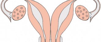
Two uteruses and two cervixes – Gynecology
Complete duplication of the uterus is a rare anomaly in the development of the female reproductive system, characterized by the formation of two uteruses with separate cervixes. In some cases, the formation of two vaginas is possible, but this pathology is extremely rare.
Pregnancy at doubling
The presence of two uteruses in a woman may not be detected until the onset of labor. In the history of medicine, there are cases of simultaneous development of a child in two uteruses, but most often the fetus is in one of them.
Possible complications of pregnancy during doubling:
- abnormal position of the fetus;
- incoordination or weakness of labor;
- postpartum bleeding (especially if the vaginal septum is ruptured);
- premature birth;
- miscarriage.
However, the development of two uteruses in a woman is more often determined during a gynecological examination or infertility treatment. The inability to conceive and bear a baby is a common occurrence when an organ is duplicated. It is typical for 50-60% of cases of atypical development.
A woman’s two uteruses can be absolutely complete; in a number of other cases, one of them turns out to be underdeveloped. With complete doubling of an organ, pregnancy may occur in any of them.
If there is one developed and the second rudimentary pregnancy occurs in a full-fledged uterus. The development of the fetus in rudimentary form is considered an ectopic pregnancy and becomes a threat to the woman’s health.
A defective organ may rupture and the fetus may enter the abdominal cavity.
Factors in the development of the anomaly
A woman's two uteruses are formed during intrauterine development. The reason is the lack of fusion of the two initially developing uterine cavities. Failure of proper formation is caused by the unfavorable factors presented below.
- Lack of a healthy lifestyle culture among parents (especially the mother). First of all, this concerns habits and living conditions that lead to chronic intoxication of the body. Addiction to alcohol, nicotine or energy drinks also leads to constant intoxication, leading to negative consequences.
- Infections of the reproductive system suffered during pregnancy or immediately before conception. It is extremely important to follow the recommendations of specialists when planning a pregnancy, since after treatment of some infections a long recovery period is required. At this time, conception is not recommended.
- Situations that promote the release of stress hormones (cortisol, adrenaline, prolactin). They have an extremely negative effect on internal processes in the body. The best method to combat this factor is a good mood and an optimistic outlook on life. During pregnancy, it is especially important to monitor your emotional background.
- Hormonal imbalances. Hormones regulate the normal functioning of all human organs and systems, including the processes of formation of the unborn baby. Disruption of the endocrine system provokes developmental pathologies, including uterine duplication. The following pathologies can cause hormonal imbalance: diabetes mellitus, diseases of the thyroid gland, ovaries and adrenal glands.
- Malnutrition. Depletion of the diet in vitamins or minerals leads to various changes in the body, including the development of a number of anomalies.
The listed factors are not specific. In addition to the development of two uteruses with two cervixes and (rarely) two vaginas, they can cause any pathology of fetal formation or provoke diseases in the woman.
Important! A healthy lifestyle and the elimination of harmful factors that affect health are the best basis for the development and birth of a healthy baby.
Symptoms of doubling
A woman with two uteruses may never feel pathology in herself. Situations where such a structure does not cause negative feelings and does not interfere with the birth of a healthy child are considered the norm. Menstruation occurs simultaneously in both uteruses. However, various manifestations are possible:
- 3 months after the onset of the first menstruation, a pronounced feeling of fullness in the lower abdomen occurs.
- Brown or bloody spotting may appear in the middle of the cycle. The symptom occurs when there is a fistulous tract between two uteruses. This opening is located in the septum that separates the two vaginas.
- Penetration of infection into the uterine cavity through a fistulous tract can be manifested by the discharge of blood with pus, which becomes permanent.
- The manifestation of the presence of a double uterus during childbirth is characterized by a decrease or discoordination of labor. Heavy postpartum bleeding is also possible.
- Infertility.
- Inability to carry a child to term, premature birth or miscarriage.
- Cycle disturbances, heavy or scanty menstrual flow.
- Periodically occurring cramping pain in the lower abdomen.
The listed symptoms do not always indicate directly a bifurcation of the organ, but only indicate a deviation in the health of the reproductive system. Given the rarity of the pathology (about one case in four million women), doctors identify this feature only after conducting a thorough examination.
The development of 2 uteruses with 2 cervixes may not always have manifestations. About 50% of women do not feel the peculiarity of the structure and learn about the anomaly during an ultrasound examination of the pelvic organs. However, even ultrasound does not always determine pathology. In the absence of symptoms, doubling is considered normal and does not require treatment.
Diagnostics
The diagnosis of doubling of the main reproductive organ is made on the basis of a complete gynecological examination, which consists of the following:
- collection of complaints and inspection;
- Ultrasound of the pelvis;
- MRI of the pelvic organs;
- hysteroscopy;
- colposcopy;
- Ultrasound examination of the kidneys.
An experienced gynecologist can identify or suspect a developmental anomaly (if there are two cervixes) during a routine gynecological examination.
Treatment
If the reproductive system is functioning normally, no treatment is required. During pregnancy, control over the condition of the uterus and fetal development increases.
However, if the menstrual cycle is disrupted, pain occurs, or infertility develops, surgical intervention is necessary. The type of operation is selected taking into account the structure of the organs, the development of both halves of the uterus and the general health of the woman.
If the outflow of menstrual fluid is disrupted, severe pain syndrome (algomenorrhea) is observed. This complication occurs in adolescence after the onset of menstruation and requires surgery.
The surgeon excises the septum between the uteri, forms a single cavity and sanitizes the vagina. This type of intervention is called metroplasty (formation of one uterine cavity from two existing ones).
In the absence of serious disorders, when only a disruption of the menstrual cycle is observed, hormonal therapy is used. No surgery required.
Important! Duplication of the uterus affects the functioning of the pelvic organs. Treatment of organ duplication must be carried out under the supervision of a nephrologist and urologist.
Source:
Two uteruses in women and pregnancy, diagnosis, treatment, risks
In obstetric practice, there are sometimes unique cases of a woman having two uteruses. This natural anomaly causes concern among owners. Having received such a diagnosis, a woman begins to worry about how to get pregnant if there are 2 uteruses.
This feature of the development of the organ does not contradict its main function - childbirth. Obstetricians know a sufficient number of cases of successful motherhood with a similar pathology.
What to do if a double uterus is diagnosed? How is pregnancy progressing? What features should you pay attention to? Detailed information is below in the article.
Features of the female body with a bifurcated uterus
The pathology begins during the period of embryonic development. There are many reasons for the incorrect placement of an organ.
In the first stages, under the influence of negative environmental factors, the embryonic uterus forms very slowly. This leads to the fact that the woman ends up with two uterine cavities. This type of pathology is the most common.
If the developmental deviation was too strong, then the uterine cervix, and even the vagina, can double in size. Most women with this pathology are not even aware of its presence.
Developmental anomalies are diagnosed during pregnancy using ultrasound examination.
The abnormal structure of the organ does not affect the menstrual cycle and egg maturation. However, pregnancy often comes with complications. Therefore, women with two uteruses require closer medical supervision.
Often such a pregnancy requires hospital stay throughout the entire period of the child's development.
Ways to treat pathology
In most cases, 2 uteri do not require special treatment. For women, this pathology does not cause discomfort. In complex forms of organ abnormality, surgical intervention may be required.
Indications for surgical treatment of a double uterus:
- acute inflammatory processes that occur in the organ cavity;
- inability to pass menstrual flow.
In such cases, the woman has the pathological organ or its paired part removed. All other cases of disease do not require such a radical solution.
Also, removal of the second uterus is used if a woman has problems conceiving. In this case, one organ is surgically formed from two uteruses. As a rule, this method solves the problem of conception.
If during the examination it is determined that one of the uteruses cannot contain a fetus, it must be removed.
Classification and reasons
The classification of this pathology is based on the doubling of certain parts of the female reproductive system. Highlight:
- complete duplication of the uterus and vagina;
- doubling of the uterine body only (bicornuate uterus);
- doubling of the body and neck.
Pathology of the structure of the uterus is often accompanied by defects in the development and functioning of the urinary system.
Reasons why women develop two uteruses:
- maternal use of steroid drugs;
- use of antibacterial dosage forms;
- excessive consumption of alcoholic beverages;
- nicotine entering the fetus’s body due to maternal smoking;
- severe forms of toxicosis and gestosis;
- hereditary and genetic predisposition;
- incorrect hormonal composition in a woman who is carrying a child;
- improper attachment of the placenta;
- viral and infectious processes in the mother’s body;
- inflammation of the female reproductive system.
The most common cause of uterine pathology is the incorrect lifestyle of the expectant mother. Alcohol, smoking, and drugs disrupt the process of formation and formation of internal organs.
As a result, a pathology in the form of two uteruses may occur.
Characteristic symptoms and diagnosis
Two uteruses in women have the following symptoms:
- pain in the lower abdomen;
- irregular menstrual cycle;
- bloody discharge from the vaginal cavity;
- purulent vaginal discharge;
- absence of menstruation;
- problems with conception and infertility;
- spontaneous miscarriage after conception has taken place;
- severe bleeding during childbirth;
- insufficient labor activity or its complete absence.
Unpleasant symptoms occur with concomitant diseases and pathologies. The presence of two uteruses, as a rule, is asymptomatic for a woman and does not cause discomfort.
Very often, this pathology is diagnosed by ultrasound during a routine examination or during pregnancy registration. This is due to the fact that the pathology, in most cases, occurs without negative manifestations.
Diagnosis of pathology
As noted above, a woman may not suspect for quite a long time that she has a double uterus. This pathology manifests itself at the beginning of sexual activity, or when problems arise with pregnancy in general. During a gynecological examination, women usually do not detect external changes in the genital organs.
Diagnosis of pathology consists of the following comprehensive measures:
- Collecting and studying anamnesis to compile a complete clinical picture of the disease.
- A thorough study of the reasons that provoke the development of the anomaly.
- Performing a gynecological examination, which helps to identify manifestations of pathology, pushing the woman to better diagnostics for an accurate diagnosis.
- Hysteroscopy.
- Vaginoscopy.
- Study of a woman's genital organs using MRI and ultrasound. These are the most commonly used methods that allow us to identify pathology in any form. Using ultrasound and MRI, it is possible to identify renal agenesis and determine the size and structure of the uterus. MRI makes it possible to determine the most optimal method of surgery used.
- Additional examination of the kidneys to avoid the development of their pathologies.
- Colposcopy to determine abnormalities in the cervix. If its doubling is detected, this method of examination allows us to identify the location and size of the reproductive organs.
When to see a doctor
Complete duplication of the uterus is mainly detected during a routine gynecological examination. It is noteworthy that about 40% of the total number of patients learn about the presence of pathology due to incorrectly prescribed treatment or due to incorrect actions of doctors: unintended removal of appendages, bougienage of the cervical canal, and appendectomy.
It is often not very easy to determine the exact cause that led to the doubling of the uterus, especially in teenage girls. Therefore, when pathology is detected and existing congenital anomalies of the reproductive system, gynecologists strongly recommend performing a full gynecological examination in order to be able to determine malformations of other organs. In most cases, the MRI method is used for this, which allows you to obtain the most complete information about the condition of all female genital organs.
Two uteruses in women and pregnancy, diagnosis, treatment, risks
Duplication of the uterus is a congenital developmental anomaly in which there are different options for dividing the main reproductive organ of a woman into 2 parts. Pathology is one of the causes of infertility and menstrual dysfunction.
The causes of abnormal development of the uterus are most often harmful factors affecting a pregnant woman when carrying a female fetus. Primary detection of the disease is possible with an ultrasound scan, but for better visualization of the malformation, an MRI of the pelvis should be done.
This is especially important against the background of habitual miscarriage, infertility and preparation for surgical intervention.
Types of uterine duplication
- Duplication of the uterus and pregnancy
- Diagnosis of uterine duplication
- MRI with double uterus
- Treatment of uterine duplication
- genetic disorders;
- complicated pregnancy in the mother (threat of miscarriage, gestosis, malnutrition, intrauterine infection);
- long-term use of toxic medications during pregnancy;
- occupational hazard in a pregnant woman;
- severe diseases of internal organs in the expectant mother.
Causes of the anomaly
Duplication of the uterus and other defects of the female genitourinary organs occur in no more than 1% of women (0.3-0.9%).
The reproductive and urinary system is formed in utero from one embryonic germ, which can cause the formation of several types of defects (a double uterus is often combined with a double kidney or other types of congenital pathology of the urinary system). Abnormal intrauterine development of the genitourinary organs in a girl occurs against the background of the following causative factors:
It is often impossible to accurately identify the main cause that led to the occurrence of intrauterine pathology in a girl.
Therefore, in all cases when a girl or woman is found to have congenital anomalies in the reproductive system, it is necessary to conduct a full examination to identify malformations of neighboring organs.
It is optimal to use the MRI method for this, with which you can obtain maximum useful information about all genitourinary organs.
What is a bicornuate uterus?
Duplication of the uterus and vagina is a type of structure in which the organ is divided by a septum into two chambers. Depending on the severity of anatomical changes, physiological processes in the organ, including pregnancy, can occur in different ways.
The causes of such disorders may be viral and bacterial diseases, pharmacological agents, addictions, and occupational hazards.
The influence of teratogenic factors is especially dangerous during the critical period of fetal development (8-16 weeks of gestation).
Uterus doubling options
Malformation of the uterus is almost always combined with pathology of the cervix and vagina. There are several possible types of congenital uterine anomalies.
| Variant of malformation | Description |
| Full doubling | 2 uteruses and 2 vaginas are completely separated and separated from each other |
| Incomplete doubling | 2 isolated parts of the reproductive organs (uterus and vagina) in a certain area are connected by a fibromuscular septum |
| Complete doubling with one vagina | 2 uteruses, 2 cervixes and 1 vagina |
| Double organ with one cervix and vagina | 2 uteruses, but the cervix and vagina are presented in 1 version |
| Duplication of the uterus with a rudimentary horn | One half of the organ looks normal, and the second is underdeveloped and is represented by a defective cavity |
| Two-horned | The uterine cavity is deformed in the upper part, partially dividing the organ into 2 halves |
| Saddle | The fundus of the uterus is moderately deformed without dividing the organ into 2 parts |
| Internal partition | A thin fibrous septum inside the uterus separates the organ completely or partially |
It is not always the case that a young woman experiences typical symptoms and manifestations, so a congenital defect in the reproductive system is discovered by chance at the stage of preconception preparation or during an examination for infertility.
The asymptomatic course of the disease is especially common with erased and unexpressed variants of doubling (bicornuate, saddle-shaped).
It is much worse if the anomaly of the genital organs is combined with a delay in the outflow of menstrual blood: in this case, typical symptoms will become the basis for an accurate diagnosis.
Symptoms of congenital pathology
Manifestations of congenital pathology depend on the form of uterine duplication. There are 2 groups of defects possible:
No retention of menstrual blood in the uterine cavity
The vast majority of congenital anomalies of the uterus are represented by defects in which a woman has normal periods. With a regular cycle, women who do not have an intimate life will not have any symptoms or unpleasant manifestations of congenital pathology.
With the onset of sexual activity, problems may arise associated with the act of love, which happens with a double vagina against the background of complete doubling. The main symptoms occur in women who want to give birth to a baby.
With uterine abnormalities without menstrual problems, the following manifestations are possible:
- threat of miscarriage at any stage of pregnancy;
- spontaneous abortion in the early stages;
- late miscarriages;
- habitual miscarriage;
- premature birth;
- infertility.
In all cases of prematurity or in the absence of a desired pregnancy, it is necessary to do a full examination, starting with safe and highly informative examination methods (ultrasound and MRI).
Duplication of the uterus is an unpleasant malformation of organs
Defects with delayed blood outflow
The lack of communication between one part of the uterus and the cervix and vagina is the reason for the retention of menstrual blood and the appearance of the following symptoms:
- abdominal pain associated with menstrual periods;
- detection of a tumor-like formation gradually increasing in size in the lower abdomen.
Typically, such symptoms occur when the uterus is doubled with a functioning rudimentary horn. If the underdeveloped area of the double organ communicates with the cervix, then the following problems are possible:
- bleeding before and after menstruation;
- heavy periods;
- ectopic pregnancy.
Any types of defects can lead to the formation of genital endometriosis with typical symptoms.
Duplication of the uterus and pregnancy
Symptoms
The presence of two separate uteruses and vaginas may not cause clinical manifestations and can be diagnosed during a routine gynecological examination, ultrasound, or surgery.
In the case of complete doubling of the uterus and vagina in combination with aplasia or atresia of one vaginal cavity, 3-6 months after the first menstruation, a picture of hematocolpos, hematometra, hematosalpinx develops on the side of the separate cavity.
In this case, the symptoms are characterized by severe bursting pain in the lower abdomen, which cannot be relieved by antispasmodics and painkillers. With a saddle uterus and intrauterine septum, clinical manifestations in adolescence are usually absent.
If there is a fistula tract in the intervaginal septum, menstrual blood can partially flow through the full vagina. When a secondary infection occurs, pyocolpos, pyometra, and pyosalpinx are often formed. Patients with a fistula opening in the intervaginal septum experience constant bleeding or purulent leucorrhoea from the genital tract.
The combination of uterine doubling with ovarian hypofunction may be accompanied by algodismenorrhea, amenorrhea, and infertility. If one or two uterine cavities are complete, pregnancy is possible, but usually occurs with the threat of spontaneous abortion and premature birth. Childbirth with a double uterus is often complicated by incoordination or weakness of labor, heavy postpartum bleeding, and lochiometer.
Duplication of the uterus and pregnancy
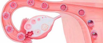
The desired conception against the background of complete doubling of the reproductive organ is quite possible. Pregnancy begins on the side where the egg is released from the ovary. The beginning of pregnancy may be accompanied by the following symptoms:
- menstrual-like reaction during pregnancy (a woman experiences scanty bleeding on the days of her expected period);
- brown discharge in the first months of pregnancy (rejection of the endometrium from the part of the uterus where there is no fertilized egg);
- nagging pain in the abdomen.
All these manifestations are similar to typical symptoms of a threatened miscarriage, so the doctor will prescribe maintenance treatment or refer you to the hospital. If no serious problems arise in the first stages of fetal development, then the woman, with constant monitoring and treatment, carries the fetus to term and gives birth to a healthy baby.
In difficult cases of infertility associated with uterine duplication, IVF is performed. After a full examination, including MRI, the fertilized “in vitro” egg is implanted in that part of the future fetal sac where conditions for gestation are most optimal.
Fetal development with incomplete duplication of the uterus
Diagnosis of uterine duplication
The main methods for identifying various types of abnormal structure of the reproductive system are the following studies:
- transvaginal ultrasound;
- hysteroscopy;
- MRI;
- laparoscopy.
At the first stage of the examination, preference is given to non-traumatic methods - ultrasound and MRI. The doctor can easily detect a saddle-shaped and bicornuate uterus with an ultrasound scan. Endoscopic techniques are optimal when combining diagnosis and treatment.
Visual examination of the inner surface of the uterus (hysteroscopy) allows you to detect the septum and immediately remove the developmental anomaly. External examination of the uterus during laparoscopy is combined with an operation to remove a non-functioning part of the organ in the presence of a rudimentary horn.
What will a tomography show when the uterus is doubled?
MRI with double uterus
Magnetic resonance imaging allows you to obtain a three-dimensional image of the reproductive and urinary organs. MRI examination is indicated in the following cases:
Source: https://sakhbmc.ru/ginekologiya/beremennost-pri-udvoenii-matki.html
Treatment of pathology
If a duplication of the uterus, irregular menstruation, or partial aplasia of the vagina is diagnosed, surgery should be performed. The surgeon makes an incision in the vaginal walls, creating a kind of thread between the uterine cavities, ensuring the outflow of hematocolpos. The patient is prescribed vaginal sanitation.
Laparoscopic examination is performed to clarify the location of organs. It is very important to remove existing accumulations of blood in the uterus and its tubes, and to examine the entire peritoneum.
Extirpation of the rudimental uterus through laparoscopy helps to determine the additional formation of a closed horn. The procedure allows you to save the fallopian tube, as well as the ovary.
If an intrauterine septum forms, as well as with problematic functioning of the reproductive system, the patient may be prescribed meteroplasty. If a doubling of the uterus with bilateral aplasia is detected, abdominal colpopoiesis and colpoelongation are used.
Examination by specialists
If, in addition to the disturbed structure of the genital organs, a woman was found to have abnormal functioning of the bladder and kidneys, then additional therapy should be prescribed by a nephrologist, as well as a urologist. For each woman, the doctor must choose his own special method of treating duplication, since the reproductive organs of different patients have a distinctive anatomical structure.
As it became clear, complete doubling of the uterus and pregnancy are compatible things. The main thing is to detect the problem in a timely manner, follow all recommendations, and complete the full course of treatment.