A formation called anechoic is nothing more than an inclusion that has arisen in any organ, which does not reflect ultrasound rays. You should not treat this categorically, since this phenomenon is not only a pathology, but also a variant of the norm. The organ itself in which these abnormalities are visualized plays a certain role in making a diagnosis.
The exact definition of the term “anechoic” is “not capable of reflecting sound.” In the ultrasound image, the inclusions that have arisen will be represented by dark spots. This is often how fluid formation (cysts) manifests itself.
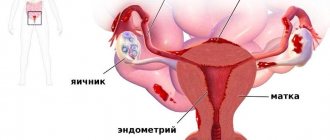
The ovary and its structure
Depending on the time of the menstrual cycle, the anechoic formation in the ovary can represent completely different structures. It should be remembered that not all of them are pathologies.
Physiological inclusions
At the end of menstruation, the anechoic formation that appears in the ovary may well be an enlarging follicle. The characteristics of this structure are as follows:
- Round form.
- The average size is from 7 to 12mm.
- It can be presented in several copies, the maximum size is up to 30 mm.
After ovulation, the inclusion that does not allow ultrasound waves to pass through can be the corpus luteum. If during this period a woman notices the presence of menstrual delay, you should worry about conducting a pregnancy test, which can be done by ultrasound at home. If the result is positive, the anechoic formation in the ovary is the luteal body of pregnancy. And even though the fetus is not yet visualized, this anechoic inclusion already creates the necessary environment for it to fully develop. After 12-16 weeks of pregnancy, the placenta will do this.
Options for deviations from the norm
In addition to the follicle and corpus luteum, the dark spot on the ultrasound image may well be an anechoic ovarian cyst. Moreover, this deviation occurs both against the background of a pathological nature and due to excessive functional activity of the organ (often not dangerous).
The classification of cysts is as follows:
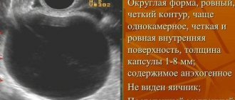
- Follicular. Can be diagnosed in the second half of the menstrual cycle. The cyst is avascular (no blood supply), about 3 cm in diameter, round in shape. A homogeneous anechoic structure, surrounded throughout its entire volume by a thin capsule. A variant of the norm is self-elimination for a maximum of 3 cycles.
- Corpus luteum cyst, which occurs after ovulation. The parameters are similar to the follicular one, resolution is carried out in approximately the same time frame.
- Cysts requiring surgical intervention (endometrioid, dermoid); malignant formations. There are two-chamber or multi-chamber varieties (cystoma), growths on the walls and echo-positive inclusions.
It is unlikely that it will be possible to determine the nature of the formation that has arisen and its absolutely exact location. Having identified fluid formations in the immediate vicinity of the ovary, the specialist will not rule out a possible cyst.
Characteristics of anechoic inclusions
Based on the histological structure and mechanism of formation, gynecologists distinguish two types of cysts:
- Retention (Nabothian) cysts . Small formations containing liquid inside. As a rule, this is the secretion of the mucous glands of the cervix and cervical canal. They do not cause discomfort to a woman; they are most often discovered by chance during a routine gynecological examination. Any symptoms appear only when the inclusion is infected.
- Endometrioid cysts . They are cells of the lining of the uterus. The growth of such formation is hormone dependent. During the menstrual cycle, such a cyst goes through all stages of transformation characteristic of the endometrium. They appear as spotting 3–7 days before and after the end of menstruation.
Breast defects
Having diagnosed an anechoic formation in the mammary gland, the doctor assumes the presence of a cavity with transparent contents (probably liquid). Often such a formation is a cyst. However, a galactocele, a cavity filled with breast milk characteristic of a woman during lactation, falls under the same description.
An ordinary simple cyst has a homogeneous structure that does not reflect ultrasound waves. With complex variations, a hyperechoic formation may be observed in the cavity. However, both options are susceptible to the development of cancer cells in them. On this side, uneven and deformed cysts and cysts with various inclusions pose a great danger.
A competent mammologist can determine the nature and nature of the occurrence of any of the formations (hyper-, isoechoic or anechoic). Most likely, this will require not only an examination and an ultrasound image, but also the results of a biopsy.
Ultrasound diagnostics of anechoic formations
The examination of the cervix is carried out in conjunction with other organs of the female genital area. The examination is carried out with a transabdominal and vaginal location of the sensor. In the first case, the doctor moves the scanner along the anterior abdominal wall in the groin area, in the second, a special sensor is used, which is inserted into the patient’s vagina.
It is recommended to perform a transabdominal ultrasound with a full bladder. To do this, you need to drink about a liter of still water an hour before the procedure. On the contrary, a vaginal examination is performed with an empty bladder.
The scanner sensor is lubricated with a special contact gel, eliminating air gaps between the skin and the device. Before insertion, a disposable condom is put on the vaginal sensor, which is also lubricated with a special gel.
The timing of diagnosis depends on the menstrual cycle - the most favorable timing is from 5 to 10 days from the start of menstruation. Ultrasound examination is painless. Pain is possible during acute and chronic inflammatory processes in organs. The duration of diagnostics does not exceed 20 minutes.
Deformation of the thyroid gland structure
When anechoic formations are diagnosed in a specified organ, it may be:
- Pseudocyst. The inclusion is not round in shape, but has a flocculent structure. Its walls are formed not by epithelium, but by gland tissue.
- True cyst. Quite a rare occurrence for the thyroid gland. It has a round shape, neat, even outlines, with the effect of dorsal reinforcement.
- Benign formation (adenoma). Depending on the cellular composition, the contents may be anechoic or hyperechoic.
- Anechoic avascular formation. Often these are colloid cysts, which have a rather low density. They appear due to the lack of a sufficient amount of iodine.
A formation in the thyroid gland can be detected by donating blood for hormones or performing a biopsy.
Deformation of the uterine structure
If an anechoic formation is detected in the uterine cavity, all possible scenarios should be considered:
- Benign tumor of the uterus (leiomyoma).
- Fluid from a ruptured follicle. This is the norm if the pathology is detected during ovulation or within 2 days after it.
- Malnutrition of myomatous nodes.
- An emerging hematoma. This is relevant when an anechoic formation in the uterus is detected in the suture area.
- Pregnancy or imminent menstruation. You can find out by performing a vaginal ultrasound after 2-3 days.
If these inclusions are found in the cervix, this is:
- Endocervical cyst.
- Nabothian gland cyst. It is a kind of cavity containing mucous secretion, which is formed when the excretory ducts are blocked. Arises as a consequence of self-medication of ectopia, erosions, etc.
- Endometrioid cyst (the walls of the detected inclusion are thickened).
- Cervical cancer. Characterized by the presence of heterogeneous inclusions with varying echogenicity. The neck thickens and changes shape.
In patients who have given birth, the detection of anechoic formations is the norm, but only with sizes up to 5 mm.
Causes of cystic formations
Cervical cysts occur in 15–18% of women of reproductive age. There is a whole range of reasons leading to their development. The main ones are the following:
- Impaired patency of the excretory canals of the glands . They are located in the epithelial layer of the cervical canal. The outflow of secretion becomes impossible; it accumulates in the gland duct and, expanding it, leads to the formation of a cyst.
- Chronic inflammatory diseases . Active division of epithelial cells in the area of inflammation, changes in the nature of mucous secretions, and a large number of desquamated epithelial cells lead to blockage of the glands and the development of cystic formations.
- Ectopic uterine endometrium . In this condition, cells of the uterine mucosa migrate to unusual places. This is the main reason for the formation of endometriotic formations of the cervix.
Risk factors for the development of pathology are traumatic damage to the mucous membrane of the cervix and cervical canal, occurring in the following cases:
- childbirth;
- abortions;
- diagnostic and therapeutic curettage;
- installation of an intrauterine device;
- hysteroscopy.
Hormonal changes in the body during menopause and chronic cervicitis also contribute to the occurrence of cystic formations.
Pregnancy period
In the fetus during the prenatal period, the detected formation is often a cyst, but its location is also important. After childbirth, these pathologies are practically not confirmed.
During pregnancy, the echo-negative structure is:
- Luteal or follicular cyst, if located in the ovary.
- Benign fluid formation.
- Fertilized egg.
In the latter case, detection is carried out at a period of 5-6 weeks; the formation is located in the upper part of the uterus and has a hyperechoic rim.
Methods of treating pathological processes
In the absence of evidence for the need to start treatment right away (ovary with an anechoic inclusion like polycystic disease, etc., small fibroids, uterine cyst), dynamic observation under the supervision of a gynecologist is indicated. If progression of the pathological process is detected over several weeks or months, specific treatment is prescribed.
Most of these diseases are treated surgically.
Laparoscopy
Cancerous tumors are excised completely or subtotally. The doctor removes as much of the changed tissue as possible to prevent further growth. A course of chemotherapy and radiation treatment is carried out to consolidate the achieved result.
Uterine fibroids are also subject to excision if they are of sufficient size. Although it is not prone to develop into cancer, it creates a mass effect and causes a lot of discomfort to the patient. In some cases, treatment is not necessary, if the neoplasia is small and does not progress, there is no need to do anything.
Cysts require drainage and puncture to pump out fluid. This technique does not always give the desired result. A relapse is likely within a couple of years or even faster. Therefore, they often resort to endoscopic removal of fibrous capsules along with the liquid itself. Multiple anechoic inclusions on the cervix allow their removal through natural anatomical passages without punctures or laparoscopic access.
Endometriosis is also treated by curettage. This is a traumatic procedure; it involves removing the top layer of the endometrium, the inner lining of the uterus. It is carried out regularly according to indications.
In some cases, a medication approach is more effective. Hormonal drugs are used, for example, for fibroids in the early stages, multifollicular ovaries.
Specialists: gynecologist, oncologist, endocrinologist. It is possible for specialists of mixed profiles to participate in therapy.
Kidney deformation
The identified anechoic formation in the kidney is often a cyst. It can be classified as follows:
- Polycystic disease. Characteristic of both organs. The kidneys are enlarged, the parenchyma is difficult to determine.
- Secondary cysts. Round in shape, localized near the scar area, the internal echo structure is changed. They appear against the background of inflammation.
- Perinephric hematoma. The organ has a familiar shape and outline; there is an area of hypoechoic parenchyma.
- Cystic carcinoma. A site with an uneven contour and mixed components.
- Abscesses. The outlines are blurred, the vessels cannot be visualized. The renal pelvis has thickened walls (more than 2 mm).
In addition to these, there are also simple cysts that have a clear round shape. They are characterized by anechoicity and thin walls. Elderly people are often susceptible to this type of inclusion.
Liver deformity
As in the case of the kidneys, the foreign structure is almost always represented by a cyst.
- An hydatid cyst is a round formation characterized by echogenic walls and the presence of calcifications inside.
- Hepatic artery aneurysm. The formation is subject to pulsation, echo-negative.
A simple variation of the cyst is characterized by septations, an oval or round shape, casting shadows along the contour.