Exposure of a tumor to ionizing rays can achieve a positive effect, since tumor cells are quite sensitive. Radiation therapy causes virtually no damage to healthy cells, even after removal of the uterus and appendages. This is the most gentle method, unlike surgical intervention, which is carried out everywhere today and the consequences are minimal. Radiation therapy after removal of the uterus is one of the most effective today.
Radiation is often carried out in combination with chemotherapy and is indicated for use at any stage of development of the oncological process. The radiation therapy method can be prescribed after removal of the uterus and appendage. While surgery may be completely ineffective.
Radiation hysterectomy is mainly carried out after surgery in order to eliminate the remaining, other abnormal structures in the structure of cells in uterine cancer in women. The basis of the radiation therapy method is the therapeutic effect, despite training with ionizing rays, the harm from which is insignificant. Although this irradiation is contraindicated if women have:
- radiation sickness;
- thrombocytopenia;
- feverish condition;
- tumor disintegration;
- severe bleeding due to tumor decay;
- myocardial infarction;
- tuberculosis;
- diabetes mellitus;
- liver, kidney failure;
- stage 4 cancer;
- anemia;
- multiple metastases.
Surgery
In most cases, surgery makes it possible to completely get rid of a malignant tumor and achieve remission. Surgical treatment options depend on the type of cancer, but in general, as a rule, a malignant tumor can be removed at stages I, II and often at stage III.
In addition to radical ones - aimed at complete removal of a malignant tumor - in oncology there are the following types of operations:
- Cytoreductive - aimed at removing the maximum possible amount of tumor tissue. Cytoreduction can be complete, when no visible tumor foci remain after surgery (but micrometastases may remain), and partial, when the primary tumor is removed, but metastases remain. Optimal cytoreduction is an operation during which most of the tumor tissue is removed.
- Palliative - surgical interventions, after which tumor foci obviously remain in the body. They are performed to improve the patient's condition if radical surgery is not possible.
- Symptomatic - operations whose goal is not to remove a malignant tumor, but to combat symptoms, for example, pain or bleeding.
- Reconstructive - aimed at restoring the function and appearance of an organ after radical removal of cancer.
The development of modern surgical oncology is proceeding in some key directions. First, surgeons are increasingly seeking to perform breast-conserving interventions in cases where this can be done without increasing the risk of recurrence. For example, in the early stages of breast cancer, complete removal of the organ—mastectomy—has become less common. Scientific studies have shown that it is often possible to remove only the part of the breast in which the malignant tumor is located, and then carry out a course of radiation therapy, without increasing the risk of recurrence.
In modern oncology, minimally invasive operations are increasingly used. Many malignant tumors in the abdominal cavity can be removed laparoscopically - through small punctures instead of an incision. Robot-assisted surgery is actively used. Early-stage cancer and precancerous lesions in hollow organs can be removed without any incisions at all, during endoscopy. Minimally invasive surgery allows you to reduce tissue trauma, blood loss, and the recovery period. Patients tolerate such operations better than open interventions.
Innovative surgical techniques are also used in the treatment of malignant tumors:
- Endovascular surgery - interventions through the lumen of blood vessels. For example, intra-arterial chemotherapy is performed (injection of chemotherapy into the artery feeding a malignant tumor), embolization (introduction of microscopic particles into the vessel that block its lumen and disrupt blood flow to the tumor), chemoembolization (injection of chemotherapy and an embolic drug).
- Stenting helps restore the patency of hollow organs blocked by a malignant tumor, for example, the intestines, stomach, and bile ducts. This is an endoscopic procedure during which a stent, a tube with a mesh wall, is placed into the blocked area. At first it is folded, and after installation it expands and expands the lumen of the organ.
- Cryosurgery is the destruction of malignant tumors using low temperature.
- Laser and radio wave surgery - using a laser beam or high frequency radio waves instead of a scalpel.
- Ablation is a procedure during which a needle is inserted into the tumor site and destroyed, for example, by heating with radiofrequency energy.
- Stereotactic radiosurgery. These interventions are often referred to as “surgery without surgery.” The device generates many X-rays or gamma rays, which are focused on the tumor site and destroy it.
The Medicine 24/7 clinic performs almost all types of modern surgical interventions in oncology, in operating rooms equipped with modern equipment from leading manufacturers.
Request a call back. We work around the clock
Intimate life after cervical cancer
If radiation therapy was successful and without complications, then many women undergo rehabilitation after it and continue to live a full intimate life. However, sometimes problems may arise in this regard, which can negatively affect not only health, but also sex.
A full intimate life is possible under the following conditions:
- if a woman has a preserved vagina. If the operation does not affect the uterus, then the woman will be able not only to live an intimate life, but even to become pregnant, carry and give birth to a child;
- complete removal of the uterus. A woman's libido is fully preserved if her ovaries are preserved. Over time, sex life will be completely restored and return to normal;
- complete removal of the organs of the reproductive system, as well as the ovaries. To restore a woman’s health and hormonal levels, special hormonal treatment is used. Doctors also perform some plastic procedures.
Chemotherapy
Chemotherapy drugs are substances that are toxic to rapidly multiplying tumor cells. There are different groups of chemotherapy drugs that have different mechanisms of action. Treatment is usually carried out with two or more chemotherapy drugs with different mechanisms of action. Combinations are selected in accordance with treatment protocols. These documents are based on large clinical studies. International treatment protocols are regularly updated. Doctors at the Medicine 24/7 clinic work in accordance with the latest versions.
Chemotherapy for cancer is used for different purposes:
- Neoadjuvant - performed before surgery. It helps to reduce a malignant tumor, reduce the volume of surgery, and convert inoperable cancer into operable one.
- Adjuvant - prescribed after surgery, helps destroy cancer cells remaining in the patient’s body after surgery and reduce the risk of relapse.
- In advanced stages of cancer, chemotherapy becomes one of the main treatment methods.
- Also, in later stages, chemotherapy drugs are used to combat symptoms.
Chemotherapy is given in cycles. Each cycle consists of drug administration and a break, usually lasting from 1 to 4 weeks. The course consists of several cycles. Neoadjuvant and adjuvant treatment always consists of a certain number of courses. Chemotherapy for inoperable cancer is continued indefinitely, as long as it helps and does not cause serious side effects. Over time, cancer cells develop new mutations and become resistant to treatment. At the same time, control examinations show that the tumor foci have begun to grow again. In this case, the doctor selects other drugs and prescribes second-line therapy.
In some cases, chemotherapy is prescribed along with radiation to the tumor. This treatment is called chemoradiotherapy.
When the drugs are taken as tablets or administered intravenously, the chemotherapy is called systemic chemotherapy. Antitumor agents can also be injected directly into the desired area of the body. This allows you to achieve a high concentration of the drug in the right place without the risk of serious side effects:
- Intra-arterial chemotherapy - the drug is injected into the blood vessel that feeds the malignant tumor.
- Intraperitoneal - into the abdominal cavity.
- Intrathecal - into the cerebrospinal fluid.
- Intravesical - into the bladder.
An innovative technique that combines surgery and chemotherapy is hyperthermic intraperitoneal chemotherapy (HIPEC). It is used for malignant tumors that have spread into the abdominal cavity and caused peritoneal carcinomatosis. An operation is performed during which all macroscopic tumor foci are removed, then a chemotherapy solution heated to 42 degrees is injected into the abdominal cavity. This helps destroy small tumor foci.
Radiation therapy after removal of the uterus and appendages
When conducting radiation therapy after removal of the uterus for uterine cancer, radiologists need to deliver a radiation dose to the tumor or possible sources of metastases that allows them to destroy malignant cells without affecting the viability of healthy tissue.
Radiation treatment is used in gynecological oncology for malignant tumors of the cervix and uterine body in the form of independent and combination therapy, in combination with surgical treatment and chemotherapy. This is a difficult treatment method that requires preliminary psychological and physical preparation of the patient.
Influence
Ionizing radiation is generated by special devices and preparations, it is used for therapeutic purposes, and is carried out after operations in all cases of histological confirmation of malignant neoplasms of the uterus, except for well-differentiated stage IA cancer.
Depending on the extent of the process, the cellular structure of the tumor and its differentiation, external, intracavitary and combined radiation therapy is used:
- In the preoperative period.
- After operation.
- For the purpose of castration for inoperable tumors.
Sources of therapeutic ionizing radiation are cobalt, gold, cesium, radium and its isotopes. For therapeutic purposes, short-wave radiation is used, which has the greatest penetrating ability into tissues, using special filters containing radioisotopes to protect healthy cells from the damaging effects of radiation.
Postoperative radiation therapy helps destroy remaining malignant cells, reduces metastasis, and the possibility of cancer recurrence.
In some cases, it is performed in the preoperative period to reduce the size of the tumor, during and after surgery. The sensitivity of different tumor cells to radiation varies; actively dividing cells are the most sensitive.
Remote
In remote therapy, the radiation source is located outside the body at different distances from the tumor (gamma units, betatrons, linear accelerators), and can be stationary in relation to the patient or moving at a controlled speed.
The absorbed dose of radiation, exposure time, number and shape of fields depend on the power of the device, the characteristics of the tumor and the body as a whole. For uterine cancer, rays are usually prescribed to the pelvic area in a total dose of 45-50 Gy.
The success of the therapeutic effect and the degree of manifestation of possible complications depend on the calculation of the irradiation dose, the number of sessions and the time of their implementation. These parameters are calculated individually for the entire course of treatment and separately for sessions.
The technical characteristics of the equipment, the level of training of medical personnel, the qualifications of a radiologist, and a physicist engineer play an important role in the process of radiation therapy.
Internal
With internal irradiation methods, a source of radionuclides is introduced into the patient’s cavities, tissues, or in the form of a pharmaceutical preparation orally.
Intracavitary gamma therapy involves inserting an applicator into the cavity of the affected organ, after which the correct installation is checked, the radioactive drug is placed in the applicator, creating optimal conditions for irradiation with a uniform distribution of energy around the source.
To deliver the drug directly to the tumor, endostats are used - hollow metal tubes with bends; they ensure reliable fixation of the radioisotope in a certain position in relation to the lesion. There are colpostats (for the vagina), metrastats (for the uterine cavity), metrocolpostats.
The introduction and fixation of metrastats takes place under intravenous anesthesia after dilatation of the uterine cavity, after which radioactive radiation sources are placed in them.
The dose is selected individually according to special tables, administered over three to five sessions at intervals of five to six days in specialized radiology departments in manual or mechanical mode, using special equipment. During sessions, it is recommended to administer painkillers and antispasmodics.
Preparation
In radiation therapy departments, medical documentation is drawn up for each patient, radiologists conduct a conversation with the woman and relatives - they explain the stages of treatment, possible complications, diet, and features of the treatment regimen. Before radiation therapy, the patient is examined for the presence of concomitant diseases that may complicate radiation treatment or make it impossible.
Various inflammatory diseases that can be pre-treated may be temporary obstacles to radiation treatment; the presence of cardiovascular diseases, diabetes, and dermatitis require preliminary preparation of the patient.
Stages
Radiation begins only when the malignant nature of the tumor is confirmed in the absence of contraindications and the conditions for radiation therapy are available. Preparation for irradiation consists of several stages.
- Laying the patient on the therapy table in a motionless position.
- Installation of the radiation head of the device.
- Pointing the radiation beam at the irradiation fields induced by the marker.
- Checking the accuracy of installation.
- Location of forming devices.
- Installation of device controls.
- Checking the readiness mode for irradiation.
- Radiation session.
Preparation can take a whole day of work by a radiologist, physics engineer, or dosimetrist technician. To accurately determine all parameters, special computer programs (COSPO), ultrasound, and CT are used.
Before irradiation, the patient is informed about the advisability of using therapeutic nutrition: excluding hard-to-digest fats, smoked meats, marinades, semi-finished products, alcohol, nicotine, and spices from food.
Diet
Food should be easily digestible, energy-rich, and in the form of puree. Preference is given to lean meats, fermented milk products, vegetables, fruits, cereals, and jelly. Long walks in the fresh air and increased water regime (still mineral water, juices, compotes, fruit drinks) are mandatory. Oncologists recommend yoga and breathing exercises.
The woman is warned about the need to maintain the markings of the irradiation fields to control the styling. Warm water is used for hygiene procedures during radiation therapy. The use of gels and creams is agreed with the doctor.
Contraindications
Radiation treatment has a damaging effect not only on malignant cells, the body is subjected to powerful stress against the background of the disease, changes occur at the level of cells, tissues, organs and systems.
Radiologists must be confident that the treatment will help defeat the disease, the patient will be able to withstand it, and it will not kill her. A number of pathological conditions make it impossible to use radiation therapy methods, these include:
- Changes in blood composition in the form of a decrease in the number of lymphocytes, leukocytes, erythrocytes, platelets.
- Cachexia.
- Acute inflammatory, purulent and infectious diseases, accompanied by an increase in body temperature.
- Diseases of the central nervous system.
- Kidney diseases.
- Severe cardiovascular pathology.
- The presence on the skin of supposed irradiation fields of wounds, skin diseases, allergic manifestations, pustules, local radiation reactions from previous irradiations.
- The state of the tumor before decay, decay.
- Sepsis.
- Active forms of tuberculosis.
If the patient suffers from chronic diseases that allow starting therapy, oncologists provide treatment against the background of radiation and recommend vitamins, enzymes, and antioxidants. Treatment of early radiation reactions is mandatory. Despite modern medical technologies, it is not possible to completely avoid complications during radiation therapy.
Complications
The frequency of severe complications after irradiation tends to decrease, but organs such as the intestines, urinary system, and skin are sensitive to radiation and are susceptible to the following pathological processes:
- Enterocolitis.
- Ulcerative rectosigmoiditis.
- Rectits.
- Recto-vaginal fistulas.
- Radiation cystitis.
- Vesicovaginal fistulas.
- Ureteral strictures.
- Fibrosis, skin atrophy, ulceration of individual areas.
Clinically, this is manifested by nausea, vomiting, flatulence, pain along the intestines, blood in the stool, increased frequency of stools, pain when urinating, increased frequency, urge, and blood in the urine. Peeling, tissue hardening, pigmentation, and ulceration occur on the skin. These reactions are usually called early radiation complications.
Treatment
To treat skin complications, fatty creams with methyluracil, vitamins, and corticosteroid hormones are used. To reduce the manifestations of intestinal inflammation, cleansing enemas with chamomile are prescribed for a week, after which they move on to oil microenemas.
For the same purpose, rectal suppositories with methyluracil and novocaine, platyphylline and prednisolone, anesthesin are used. Diarrhea is treated with sorbents, Imodium, astringent herbal mixtures, enzymes, and probiotics.
For nausea, antinausea drugs are used (Cerucal, Tropisetron). Radiation cystitis is treated by introducing medicinal mixtures into the bladder and using antibiotics. Small fistulas (up to 1 cm) resulting from radiation treatment close on their own within a year; larger diameter fistulas are treated surgically.
During treatment, the body temperature rises to low-grade levels, due to the absorption of destroyed malignant cells. If fever and intense pain occur, radiologists may recommend stopping radiation therapy. Radiation lymphostasis of the lower extremities is eliminated microsurgically, restoring the lymph outflow pathways.
Forecast
Modern radiation therapy, carried out after removal of the uterus and appendages, with optimal methods, does not lead to severe complications from surrounding tissues and the body as a whole.
The success of radiation treatment after removal of malignant tumors of the uterus depends on many factors - the structure of the cells of the formation, the stage of the process, the age of the woman, the chosen treatment method and the correct administration of radiation.
In the early stages of the disease, complete healing is possible. In later cases, with adequate treatment, the five-year survival rate can be ninety percent of all cases.
Source: https://uterus2.ru/disease/luchevaya-terapiya-posle-udaleniya-matki-s-pridatkami.html
Targeted therapy
Targeted drugs differ from classical chemotherapy drugs by having a more targeted effect. Each of them blocks a specific “target” - a molecule that cancer cells need to maintain vital processes, reproduce, and evade aggression from the immune system. Targeted drugs can be effective when chemotherapy fails and cause fewer side effects.
Some examples of targeted therapy for various cancers:
- of breast cancer, many HER2 protein molecules are present on the surface of tumor cells. When activated, this protein stimulates cell reproduction. It can be blocked with some targeted drugs, particularly trastuzumab (Herceptin).
- Some malignant tumors express large amounts of the EGFR protein, the epidermal growth factor receptor. It also stimulates the proliferation of tumor cells. For example, this situation occurs with lung and colon cancer. Such patients are prescribed EGFR blockers.
- VEGF inhibitors block a protein (vascular endothelial growth factor) that promotes the growth of new blood vessels that feed the malignant tumor.
- In melanoma, half of the cases have a mutation in the BRAF gene. Currently, several drugs from the group of BRAF inhibitors are used for stages III and IV of the disease.
Radiation therapy after removal of the uterus for oncology
Surgery to remove a cancerous uterus is often supplemented with radiation therapy. Ionizing rays do not harm healthy structures. Radiation therapy after removal of the uterus and appendages is considered a gentle way to destroy remaining atypical structures and metastases. The procedure can be supplemented with chemotherapy and is prescribed at any stage of tumor development.
Why is it used and how is it done?
Radiation therapy is indicated in the following cases:
- to remove abnormal structures at the initial stages of the oncological process;
- for the comprehensive fight against uterine cancer;
- after removal of the uterus to prevent relapse of the disease;
- in advanced stages of uterine cancer, when surgery does not bring results.
Irradiation leads to disruption of the functioning of the ovaries, which causes premature cessation of menstruation. Therefore, radiation treatment of patients is carried out with caution. In some cases, the ovaries are moved from the irradiated area to another location before the intervention. This reduces the risk of injury during the procedure.
The operation is performed sequentially:
- The patient lies down on a medical table, which has the ability to move in different directions. It is important that the woman remains motionless during the session. Otherwise, the rays will not reach the tumor and can damage healthy tissue.
- Women who cannot stay in one position for a long time are secured with special belts.
- After the last radiation therapy session, the patient undergoes additional diagnostic tests.
- The doctor advises the patient about the features of the recovery period and gives recommendations for correcting nutrition and lifestyle.
During the procedure, women do not feel pain or discomfort
Immunotherapy
Immunotherapy is a cancer treatment method that helps the immune system recognize and destroy malignant tumors. There are several types of immunotherapy, the most widely used in oncology are two: cytokines and checkpoint inhibitors.
Cytokines are substances that stimulate an immune response. Interferon-alpha and interleukin-2 are used to treat cancer. They are prescribed to patients with kidney cancer and melanoma. Interferon-alpha is also used for hairy cell leukemia, chronic myeloid leukemia, follicular non-Hodgkin's lymphoma, cutaneous T-cell lymphoma, and Kaposi's sarcoma.
Checkpoint inhibitors are the most innovative type of immunotherapy. These immunotherapies are monoclonal antibodies that block molecules that suppress the antitumor immune response. Essentially, these are targeted drugs with specific effects on the immune system. They are most often directed against the PD-1, PD-L1 or CTLA-4 checkpoints. In modern oncology, the most actively used immunodrugs from this group are ipilimumab (Yervoy), nivolumab (Opdivo), pembrolizumab (Keytruda), atezolizumab (Tecentriq), avelumab (Bavencio), durvalumab (Imfinzi).
Immunotherapy with checkpoint inhibitors, when the indications for its use are correctly determined, works much more effectively than chemotherapy and helps prolong the life of patients with advanced malignant tumors.
Hormone therapy
Hormone therapy is essentially another type of targeted therapy. It is used for hormonally sensitive tumors, in particular breast, prostate, and ovarian cancer.
Hormonal therapy for cancer is fundamentally different from that used for endocrine diseases. If a person has a malfunction of one of the endocrine glands, he is given hormonal drugs to replace the hormone that is missing or produced in insufficient quantities. This type of hormone therapy is called replacement therapy. The goal in cancer is to block the hormonal effects that promote cancer growth.
Radiation therapy
Radiation therapy for cancer, like chemotherapy, can be used before, after surgery, or in late stages as one of the main methods of treatment to slow the progression of the disease and relieve symptoms.
There are two main types of radiation therapy for cancer: external source and brachytherapy. In the first case, the procedure resembles radiography, but the radiation dose is much higher. In brachytherapy, a radiation source in the form of small particles is placed directly into the tumor tissue, next to it, or into the lumen of hollow organs.
There are modern technologies that allow very precise irradiation of a malignant tumor, practically without affecting neighboring tissues:
- Three-dimensional conformal radiation therapy uses computer technology to precisely determine the location of the tumor. The device directs radiation onto the patient’s body from different angles, resulting in radiation that most accurately replicates the shape of the malignant neoplasm.
- Intensity modulated radiation therapy The rays are directed at the body from different angles and their intensity is adjusted. This technology is used very often, especially if the tumor lesions are located near important anatomical structures, such as the spinal cord.
- Stereotactic radiation therapy also involves delivering radiation from different angles. Stereotactic surgery is a variation of this technique.
Radiation therapy for uterine cancer after surgery
Surgery to remove a cancerous uterus is often supplemented with radiation therapy. Ionizing rays do not harm healthy structures. Radiation therapy after removal of the uterus and appendages is considered a gentle way to destroy remaining atypical structures and metastases. The procedure can be supplemented with chemotherapy and is prescribed at any stage of tumor development.
Radiation therapy is indicated in the following cases:
- to remove abnormal structures at the initial stages of the oncological process;
- for the comprehensive fight against uterine cancer;
- after removal of the uterus to prevent relapse of the disease;
- in advanced stages of uterine cancer, when surgery does not bring results.
Irradiation leads to disruption of the functioning of the ovaries, which causes premature cessation of menstruation. Therefore, radiation treatment of patients is carried out with caution. In some cases, the ovaries are moved from the irradiated area to another location before the intervention. This reduces the risk of injury during the procedure.
The operation is performed sequentially:
- The patient lies down on a medical table, which has the ability to move in different directions. It is important that the woman remains motionless during the session. Otherwise, the rays will not reach the tumor and can damage healthy tissue.
- Women who cannot stay in one position for a long time are secured with special belts.
- After the last radiation therapy session, the patient undergoes additional diagnostic tests.
- The doctor advises the patient about the features of the recovery period and gives recommendations for correcting nutrition and lifestyle.
During the procedure, women do not feel pain or discomfort
Types of radiation therapy
After surgery, ionizing radiation is prescribed for distant metastases and residual cancer cells. Surgery may also be indicated if there is a high risk of recurrence of the pathology after a hysterectomy. The intervention is performed remotely, intracavitary, contact.
In the first case, the radiation appears at a certain distance from the lesion. In the contact type of procedure, the device is attached to the patient’s skin. With the intracavitary method, atypical cells are removed by introducing a special device to the lesion.
Before radiation therapy, diagnostic measures are necessary. They allow you to accurately calculate the required radiation dose. The doctor informs the patient about the consequences of the intervention and prescribes medications to be taken during the rehabilitation period.
A balanced diet and following all the specialist’s recommendations will help you recover faster after surgery.
Preparation for the procedure
The specialist describes the treatment plan in detail, calculating the radiation dose for the full course of therapy and separately for each session. The doctor determines the duration of the course of treatment and the duration of one procedure. Before starting radiation therapy, the following preparatory procedures are performed:
- At the hospital, the patient is offered disposable clothing. If a woman wants to stay in her clothes, then she should take care of some nuances:
- things should not fit the body or restrict movement;
- the jacket must have an open collar.
- If necessary, the patient is fixed to the table using belts, mattresses and other fasteners. These measures are necessary in order to completely limit the patient’s movements during the intervention.
- Healthy tissues and organs are covered with special protective blocks to protect them from radiation exposure.
- Before the procedure, the doctor can take a control photo to assess the correct positioning of the woman relative to the medical device.
The first radiation therapy session will be the longest. The duration of each subsequent procedure is gradually reduced.
Patients should adhere to several basic rules before treatment:
- do not dry your hair with a hairdryer;
- hide irradiated areas under clothing when leaving home;
- stop using cosmetics and sunscreen for a while;
- minimize physical activity;
- try to go outside after sunset;
- consume enough fluid.
10 days before treatment, the patient needs to adjust her diet. At this time, pickled foods, carbonated and alcoholic drinks, spices, and spicy dishes are completely excluded. A week before radiation therapy, doctors recommend that a woman do breathing exercises and increase rest time.
List of other preparatory activities
| Necessary measures for carrying out RT | What is it done for? |
| The presence of spacious clothing made of cotton material with a minimum number of seams. | The products will contribute to less trauma to the skin after the session. It is recommended to wear such items throughout the entire treatment period. |
| Purchasing herbal infusions that have an antiseptic and astringent effect (chamomile, oak bark, sage). | Rinsing the mouth with this medicine helps minimize the negative effects of radiotherapy on the body. |
| Replacement oral care products. | After radiotherapy, increased bleeding of the gums and mucous membranes of the mouth is observed. To reduce the risk of tissue damage, it is necessary to use brushes with soft bristles and pastes with a neutral chemical composition. |
Consequences of radiation
Radiation therapy provokes a number of negative consequences. These include:
- Intoxication of the body, manifested by weakness and nausea.
- Abnormal stool in the form of chronic diarrhea.
- Skin irritation in the irradiation area and the appearance of red rashes on it.
- Increased dryness of the vaginal mucous membranes.
Radiation therapy after removal of the uterus and appendages: effectiveness
Radiation therapy for cervical cancer involves local exposure to high doses of X-rays, as a result of which tumor cells are destroyed. Healthy cells are subject to minimal negative effects.
Radiation therapy used in the treatment of cancer of the female genital organs can be internal or external. Often these methods are used in combination. The duration of treatment depends on the stage of the disease and the general condition of the body. On average, it lasts from 5 to 8 weeks.
Radiation therapy for cervical cancer is highly effective only in the early stages of the disease. It is also included in the treatment regimen for large uterine tumors that are not amenable to surgical treatment.
Radiotherapy is also prescribed after surgery to eliminate any malignant cells remaining in the body. In such cases, it is combined with chemotherapy. After radiation therapy for cervical cancer, the ovaries stop functioning. Ovulation and production of female hormones stops.
This leads to absolute infertility. About 90 days after radiation, a woman enters early menopause. The doctor should discuss all these points with the patient before starting treatment.
There are several ways to preserve reproductive functions, for example, repositioning the ovaries into the abdominal cavity.
The operation is performed simultaneously with the removal of a malignant neoplasm, if the doctor recommends radiotherapy in the future.
It is possible to perform such surgery using the endoscopic method. In some cases, the development of early menopause cannot be avoided.
Risk factors for cervical cancer:
- Human papillomavirus
- Early onset of sexual activity (before 16 years of age)
- Early labor
- Frequent change of sexual partners
- Smoking
- Abortion
- Inflammatory diseases of the genital organs
- Long-term use of hormonal contraceptives
- Immunity disorder
- Age from 40 to 50 years (however, cervical cancer is getting younger worldwide and occurs in women aged 25 to 29 years).
Source: https://xn--201-5cd3baaocfczhn.xn--p1ai/lekarstvo/luchevaya-terapiya-pri-rake-matki-posle-operatsii.html
Photodynamic therapy
Photodynamic therapy is a method that is used to treat cancer and some other diseases. It consists of two stages. First, a special substance, a photosensitizer, is introduced into the patient’s body. It accumulates in tumor cells. Then the tumor tissue is exposed to light of a certain spectrum. As a result, the photosensitizer is activated, becomes toxic to cancer cells and destroys them.
Photodynamic therapy is used for cancer of the pancreas, bile ducts, esophagus, lungs, cancer and precancerous skin diseases.
Find out more. Request a call back
Personalized cancer therapy
For the average person, the term “cancer” is usually associated with one specific disease that can develop in different organs. In fact, no two malignant tumors are exactly alike. Each of them has its own molecular genetic characteristics. Therefore, there is no universal treatment.
Modern oncology is moving towards personalized treatment, that is, one that is selected individually for each specific patient. Genetic tests help select anticancer drugs that will be most effective in a particular case. Currently they are carried out in two cases:
- Tests for specific genetic defects help test whether a particular targeted drug will be effective. For example, before a woman with breast cancer is given trastuzumab, a HER2 test must be performed.
- Whole DNA analysis using next-generation sequencing (NGS) technology is prescribed in situations where a malignant tumor does not respond to any type of standard treatment prescribed in the protocols. Having discovered certain mutations in cancer cells, it is possible to select a combination of drugs that may be effective. Such analyzes with the selection of personalized therapy are carried out only in a few specialized laboratories in the world. Clinic Medicine 24/7 cooperates with such laboratories.
How to treat uterine cancer: radiation therapy, diet, surgical removal, chemotherapy, prevention
According to statistics, cases of female cancer are becoming more common. One of the leading places among cancer pathologies is occupied by uterine cancer, which is found mainly in 40-60 year old women.
In fact, the uterus is an organ with walls of three layers: epithelial, muscular and connective tissue. In the uterus, a malignant oncological process develops on the walls, and in the absence of proper therapy, it spreads to other organic structures.
This multi-layered nature of the uterus explains the presence of many different types of tumors, depending on location.
Is it possible to cure uterine cancer?
Of all oncological pathologies, uterine cancer ranks second in prevalence after malignant oncology.
Approximately 20-40 women out of 100 thousand suffer from cancer of this organ.
Unfortunately, in recent years, cases of uterine oncology have begun to occur in younger women, which gynecologists associate with the early onset of regular sexual relations among modern youth.
Timely contact with specialists at an early stage of the development of uterine cancer formations, in combination with an adequate therapeutic approach, can completely relieve a woman of the disease without further consequences or relapses.
If oncology is detected in the later stages, then it will not be possible to completely cure the cancer, however, following all the oncologist’s prescriptions and undergoing the prescribed procedures will help to significantly prolong the patient’s life.
In general, experts assure that uterine cancer can be successfully treated, and about 90% of patients live another 5 years or more after surgery. Therefore, today a diagnosis of uterine cancer should not be treated as a death sentence.
The choice of therapeutic approach is determined by the stage and scale of the tumor process, the speed of its spread and the depth of germination into tissue structures.
In addition, age and general health, planning a pregnancy in the future and other factors are very important.
Anticancer treatment usually focuses on:
- Surgery;
- Radiation treatment technique;
- Chemotherapy.
These techniques are fundamental, and additional methods are:
- Hormonal treatment;
- Immunotherapy;
- Diet therapy, etc.
Each of the techniques is unique and effective in its own way, and therefore requires more detailed study.
Surgery
Surgical intervention for uterine cancer is justified only at the initial stages of development of the pathological process.
In general, there are several surgical treatment options:
- Laser treatment. The essence of the technique is to influence the area of the uterus affected by the tumor process with a beam of laser beams. To carry out such an intervention, local anesthesia is sufficient, however, the procedure is effective only in the precancerous or very initial stages of the uterine cancer process;
- Cryodestruction. This procedure involves treating the tumor with liquid nitrogen, under the influence of which malignant abnormal cellular structures die. The technique is also effective only at the stage of incipient uterine cancer;
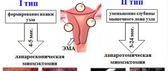
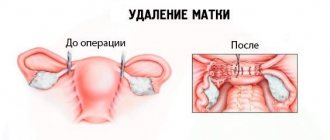
- Extription of the uterus and appendages. The procedure involves the removal of an organ and is carried out if the prognosis for oncology is favorable. If there are any risk factors, then the patient undergoes an extended hysterectomy, during which, in addition to the uterus and appendages, the tubes, cervix, lymph nodes and tissue, and part of the vagina are removed;
- Endoscopic hysterectomy allows removal without large tissue incisions, which significantly reduces the invasiveness of the method, minimizes the likelihood of postoperative complications, and reduces the duration of rehabilitation;
- Hysteroresectoscopic ablation. This is an organ-preserving surgical technique that involves cutting off the endometrial layer along with the tumor. A similar method is applicable when the tumor process is insignificant.
Most often, surgical treatment is the main therapeutic method for oncology of the uterine body.
Refusal of such treatment is appropriate only if there is no risk of further progression of the oncological process or if there is a high probability of death on the operating table or after the intervention.
Radiation therapy
If the uterine cancer process is actively progressing, surgical treatment is ineffective. In such cases, the basis of treatment is radiation therapy, which is considered more gentle than surgical treatment.
Such treatment is used at any stage of the cancer process. In addition, radiation therapy for uterine cancer is indicated in the postoperative period in order to prevent metastasis of abnormally malignant cellular structures.
Radiation therapy is contraindicated in women with uterine cancer if:
- Anemia;
- Radiation sickness;
- Thrombocytopenia;
- Disintegration of the tumor leading to bleeding;
- Fever;
- Concomitant pathological conditions such as heart attack, tuberculosis, diabetes, liver or kidney failure, etc.;
- Leukopenia;
- Multiple metastases;
- Final degrees of malignant lesions, etc.
Various radiotherapeutic techniques can be used: contact, remote or combined.
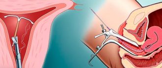
Contact radiotherapy involves internal exposure, where a catheter emitting radio waves is inserted into the vagina. With this treatment option, the surrounding healthy tissue is least exposed to the harmful effects of radiation.
With external beam (or external) radiation therapy, the radiation procedure is carried out through tissues not affected by cancer; this technique is usually used if the lesion is deep-seated. A serious disadvantage of this treatment is damage to healthy areas during treatment.
If uterine cancer is advanced and treatment is started already at the later stages of the oncological process, then combined radiotherapy is used, i.e., contact and remote irradiation methods are used.
Among the adverse reactions of such therapy are the appearance of nausea and vomiting syndrome, weakness, diarrhea, hyperemia and peeling of the skin surface, pubic baldness, etc.
Chemotherapy for endometrial tumors
The main goal of chemotherapy treatment is to reduce the parameters of the tumor and slow down its further growth in the future as much as possible.
Typically, this technique is chosen as the main therapy for stages 2, 3 and 4 of cancer.
Chemotherapy is not always used as the basis for anticancer treatment; it is often combined with other methods in order to increase the survival rate of patients.
Chemotherapy is usually used after surgery to prevent possible metastasis and relapse of the oncological process.
Most often, antitumor drugs such as:
- Cisplatin;
- Doxorubicin;
- Carboplatin.
Chemotherapy is used mainly when the use of other drugs does not provide the desired results, which is caused by a considerable number of adverse reactions of anticancer drugs such as:
- Osteoporosis. This reaction usually occurs due to the use of drugs such as Fluorouracil, Methotrexate or Cyclophosphamide and represents bone loss and weakness;
- Alopecia. Usually, after chemotherapy sessions, partial hair loss is observed, however, baldness can also be more widespread. When the use of antitumor drugs ends, the hair begins to recover;
- Anemic signs and excessive weakness of a permanent nature;
- Nausea and vomiting syndrome, diarrhea - such manifestations arise due to disturbances in the functioning of the gastrointestinal tract, however, after treatment, all signs disappear;
- Infectious processes - chemotherapeutic effects have a detrimental effect on the immune defense, depriving it of resistance to viruses and infectious pathogens.
If the cancer process has developed to stage 4, then the effectiveness of chemotherapy does not exceed 9%, since the lesions rapidly spread to the pelvic organs.
How to treat with hormone therapy?
Hormonal anticancer treatment consists of taking medications containing antiestrogens and progestins. This treatment is effective in cases where the tumor contains progesterone receptors.
If such receptors are absent, then chemotherapy treatment will be most effective.
Immunotherapy
At the initial stages of the cancer process in the uterus, immunotherapy using interferon-based drugs can be prescribed.
This substance, in addition to the strongest effect, also has antitumor properties.
The drugs administered to the patient activate the protective organic forces and direct their power to resist the tumor process.
Also, medications of biological origin, such as monoclonal antibodies or cytokines, are introduced into the body, which shut down the system that feeds the formation.
When the tumor stops growing, the malignant cancer process is blocked. This treatment does not cause any side effects and does not harm healthy tissues.
Diet
The basis of nutrition for patients with uterine cancer consists of foods with anti-cancer effects:
- Potato;
- Cabbage of all varieties;
- Onion;
- Greens, spices;
- Sprouted cereals or whole grains;
- Soy;
- Asparagus;
- Peas;
- Beet;
- Onion;
- Beans;
- Carrot;
- Fresh fruits.
The above products must be consumed fresh or cooked in a double boiler. It is recommended to replace meat with fish. You also need to eat low-fat fermented milk products. Strictly prohibited: alcohol and strong tea, smoked foods, pickles and marinades, chocolate, processed foods, fast food, etc.
It is possible to detect the cancerous process of the uterine body in its infancy only with systematic gynecological preventive medical examinations and regular visits to the gynecologist.
After starting regular sexual relations, a woman must attend an antenatal clinic annually. Only with an annual gynecological examination, vaginal smear examination, and ultrasound examination of the pelvic organs will it be possible to timely identify the presence of precancerous processes.
Their timely therapy will avoid the formation of a malignant tumor.
video about the laparoscopic method of treating uterine cancer:
Source: https://gidmed.com/onkologiya/lokalizatsiya-opuholej/reproduktivnaya-sistema/lechenie-raka-matki.html