How it manifests itself
Adenomyosis is a frequent companion to infertility and is sometimes incompatible with conception.
When an egg is fertilized, 90% of pregnancies end in the early stages due to placental abruption. When the disease occurs, the middle layer of the reproductive organ is damaged, that is, the surrounding organs are not affected by the process.
The size of the uterus increases, the functions assigned to the organ are disrupted, and ovulation processes are aggravated.
Sometimes the disease is asymptomatic and is detected after a thorough examination.
There are several signs that confirm the diagnosis in the absence of pregnancy:
- pain during sexual intercourse;
- painful periods and several days before the onset of menstruation;
- insomnia;
- headache;
- irregular bowel movements;
- blood clots in large quantities during CD;
- frequent spotting and spotting;
- weakness.
Symptoms when carrying a baby:
- nagging pain in the abdomen;
- malaise;
- change in the nature of discharge;
- blood spots;
- pain in the perineum and groin.
The clinical picture depends on the degree of pathology. With adenomyosis there are 4 of them:
- the submucosal layer was affected;
- the disease affects half of the muscle layer;
- muscle tissue is almost completely damaged;
- pronounced changes in the muscle layer.
In the gestational period, grades 1 and 2 of pathology can lead to premature birth. With grades 3 and 4 of pathology, pregnancy can end in spontaneous miscarriage or placental abruption. Increased likelihood of internal bleeding.
After birth, the child is weak, stunted in height and body weight.
Diagnostics
The diagnosis of adenomyosis in a pregnant woman is made on the basis of:
- Survey. The patient usually complains of painful and prolonged menstruation before pregnancy. And also for spotting brown-brown discharge a few days before the start of menstruation and for two to four days after cyclic bleeding.
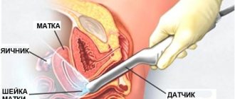
- Inspection. During a vaginal examination, the uterus is determined to be larger than expected at this stage of pregnancy. In the focal form, tuberosity may be detected. Palpation of the uterus is often painful or at least uncomfortable.
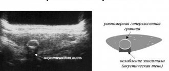
- Ultrasound examination. The discrepancy between the size of the uterus and the gestational age, increased echogenicity or thinning of muscle tissue in the areas of localization of lesions is determined.
Other examinations - magnetic resonance imaging, x-rays, hysteroscopy - are not used during pregnancy due to possible negative consequences for the fetus.
Who's at risk
Adenomyosis of the uterus is a benign neoplasm. The risk group includes women with a history of gynecological diseases and surgical interventions on the pelvic organs.
The risk of adenomyosis increases in women who have given birth and are aged 35 years or older.
Having had a cesarean section also increases the risk of developing the disease.
According to statistics, adenomyosis is registered in women of older reproductive age, but the disease can affect girls in adolescence.
Often the disease affects women:
- with hormonal imbalance;
- with an intrauterine device;
- overweight or obese;
- having vaginal sex during menstruation.
Pregnancy after treatment
In cases where the pathology was diagnosed in a timely manner, it responds well to treatment, and the woman can become pregnant in the future. If the disease is detected at the first or second stage, the prognosis is favorable, but it is still impossible to talk about a 100% recovery. Features of the course of the disease are that the menstrual cycle is disrupted and ovulation may be absent. Hormonal levels become unstable, and there is a risk of relapse.
When treating an advanced form of pathology (third and fourth stages), infertility develops in most cases. This is due to the fact that the lesions are excised surgically. At the fourth stage, they often resort to removing the ovaries and reproductive organs.
If therapy at the initial stage of development of the disease was carried out using conservative methods, then after the end of hormonal therapy there is a high chance of a healthy pregnancy.
Is it possible to get pregnant with adenomyosis?
Fusion of the endometrium and myometrium in most cases prevents pregnancy. The resulting pregnancy may be terminated in the early stages or cause bleeding.
The possibility of motherhood is directly related to the form and degree of the disease.
Stages 1 and 2 of the disease do not affect the process of fertilization and attachment of the fertilized egg to the inner lining of the uterus. Grades 3 and 4 are a big problem for the implantation of the egg.
Forms
- Diffuse. Formation of “blind pockets” and fistulas in the mucous layer. It occurs most often (about 70).
- Nodular or cystic. Formation of hemorrhages in endometrioid nodes. A brown liquid appears in the muscle tissue. It is registered in 5-10% of women.
- Mixed. The change occurs evenly with the formation of fistulas and cavity fluids.
The hysteroscopic picture is the determining factor in determining the possibility of conceiving and bearing a baby.
Is adenomyosis a death sentence for pregnancy?
Adenomyosis is one of the most common gynecological diseases in women.
According to the Ministry of Health, such a pathology occurs in every third woman in the world, and the disease can be asymptomatic in the first stages, and the patient herself may not even be aware of the presence of such a pathology. Adenomyosis can either pose a threat to women's health or not require serious treatment.
What to do to prevent illness from interfering with motherhood
A type of endometriosis negatively affects the ability to conceive and carry a pregnancy. Once a diagnosis is made, it is necessary to undergo diagnostic tests and begin treatment.
Diagnosis of the pathological process consists of:
- in collecting gynecological history;
- in a gynecological examination on a chair using mirrors;
- standard ultrasound examination and transvaginal ultrasound method;
- microscopic examination of the contents of the cervical canal;
- in sowing to establish flora;
- if necessary, hysteroscopy is performed to assess the condition of the reproductive organs and eliminate existing pathology (cysts, diffuse changes).
A laboratory blood test is performed to determine the concentration of hormones and to assess the condition of the female body: the presence of inflammation, anemia, chronic or acute diseases.
Hormone analysis helps to identify the most favorable moment for conception.
Magnetic resonance imaging and x-ray examination methods are not used to detect pathology during pregnancy.
Hysteroscopy is also not performed, because the risk of miscarriage increases.
Some specialists conduct instrumental research at their own risk, and often successfully. However, the decision remains with the patient.
Treatment and postpartum therapy
During pregnancy, treatment of adenomyosis becomes more difficult. The primary goal of doctors is to eliminate the threat of miscarriage and maintain the pregnancy. Many medications used to treat adenomyosis have a negative effect on the fetus. Therefore, therapy for pathology in pregnant women is aimed at the following:
- reduce the growth of the endometrium (androgens or progestogens are used);
- eliminate the risk of miscarriage. For this purpose, the following groups of drugs are prescribed: antispasmodics (Spazmalgon, No-Shpa);
- sedatives (Persen, Novo-Passit);
- means that improve metabolic processes (Glycine);
Adenomyosis is one of the cases of direct indication for cesarean section.
After childbirth, in some cases, adenomyosis gradually decreases. This is due to changes in a woman’s hormonal levels during the postpartum period. If the foci of the disease do not decrease, then conservative or surgical treatment is performed.
The choice of medications should be entrusted to the doctor
Conservative postpartum therapy consists of the following:
- carrying out hormonal therapy to create a state close to menopause. The drugs that are used in this case can be: androgens (Danazol);
- progestogens (Gestrinone, Dydrogesterone, Duphaston);
- GnRH analogs (Buserelin, Leuprorelin, Histrelin, Nafarelin, Goserelin);
- oral contraceptives (Zhanine, Yarina, Silhouette);
It is important to note that hormonal therapy lasts from four months to one year.
Photo gallery: medications used in complex therapy of adenomyosis
Surgical therapy
In the absence of positive dynamics after conservative treatment, doctors decide on surgical intervention. Surgery for adenomyosis is aimed at removing only the foci of the disease (hysteroscopy, laparoscopy), in advanced cases - at removing the uterus (abdominal surgery). Indications for organ removal are the following reasons:
- progressive adenomyosis in women over 40 years of age;
- diffuse adenomyosis of the third and fourth degree;
- nodular adenomyosis in combination with large fibroids;
- the threat of developing a malignant neoplasm.
Combined therapy shows the greatest effectiveness in the treatment of adenomyosis. In this case, the largest lesions are surgically removed from the myometrium, and then conservative treatment is carried out.
Treatment with traditional methods
Postpartum therapy for adenomyosis may include traditional medicine. Their effectiveness has been tested by time.
Traditional medicine is widely used in the treatment of adenomyosis
The following traditional methods are used in the treatment of adenomyosis:
- douching or taking baths from infusions of medicinal herbs (hogweed, shepherd's purse, nettle, plantain, yarrow, etc.);
- hirudotherapy (treatment with leeches);
- blue clay compresses for the groin area.
In my opinion, hirudotherapy is difficult to classify as a traditional medicine; nevertheless, leeches are considered medical, and the procedure is carried out in a hospital and under the supervision of a specialist. My friend attended several sessions with adenomyosis. According to her, each of them lasted about an hour, the pain went away and the body relaxed. However, after the session there was a feeling of severe weakness. I had to refuse to attend this procedure. And even after three years, traces of leech bites are still visible, which does not look very good aesthetically. But my friend really praised the blue clay compresses and still makes them.
It is important to note that traditional medicine also has contraindications. For example, the boron uterus is not used during breastfeeding, and hirudotherapy is contraindicated in cases of blood clotting disorders, severe anemia, and taking anticoagulants (drugs that prevent the formation of blood clots in blood vessels).
Features of pregnancy
The entire gestational period is associated with possible adverse events.
The most dangerous of them is placental abruption with subsequent death of the fetus.
- In the first trimester, the threat of gestation interruption remains. Patients receive maintenance hormone therapy. In 80% of cases, pregnancy ends before the 12th week.
- In the second trimester, the risk of spontaneous miscarriage decreases markedly, especially by week 16, when the placenta independently produces hormones. However, other problems arise. The pain increases due to the organ increasing in size. The adhesions become larger and force the fetus into an uncomfortable position and difficulties with intrauterine development.
- From the 30th week of the gestational period, subject to pronounced diffuse changes in the muscle layer, the risk of circulatory obstruction and fetoplacental insufficiency increases. Developed oxygen starvation causes developmental delays in the baby.
Adenomyosis is asymptomatic, which complicates timely diagnosis. Often diffuse changes and parts of the endothelium are found in the muscle layer (no more than 1/3).
The degree and type of pathology determines the course and outcome of pregnancy.
Some pregnant women do not experience any significant changes during pregnancy. Others are forced to be under close medical supervision.
Causes
The reasons that cause the germination of the endothelium into the muscular layer of the uterus include:
- any surgical interventions in the uterine cavity;
- physical exercise;
- Unhealthy Lifestyle;
- binge eating;
- depressive states;
- endocrine pathologies;
- diseases of the urinary system;
- arterial hypertension;
- passive lifestyle;
- long-term therapy with oral contraceptives;
- lack of sexual contact for a long time;
- immune weakness.
The disease is directly related to hormonal imbalance. Pregnancy can also cause adenomyosis.
Definition
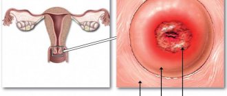
By adenomyosis, doctors mean the proliferation of endometrial cells of the inner layer of the uterus beyond its limits, which leads to complications of many natural physiological processes.
The disease is a polymorphic anomaly that can affect not only the genital area, but, going beyond it, affect the intestines.
Unfortunately, the exact causes of the disease have not yet been identified, but risk factors are:
- various genitourinary infections;
- operations on the uterus;
- trauma during childbirth;
- hormonal disorders;
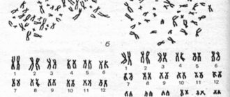
- genetic predisposition.
A history of more than two abortions increases the risk of developing the disease by more than 10 times.
What to do when diagnosed
Adenomyosis is not a death sentence for motherhood. Provided timely therapy, after treatment, the chances of becoming pregnant are quite high.
After pregnancy, a woman must strictly follow all medical recommendations.
One of the main factors in women's health and pregnancy is regular visits to the gynecologist - at least once a year. If adenomyosis is detected in the early stages, the diagnosis does not become a cause of female infertility, and after complex treatment, the desired pregnancy will occur.