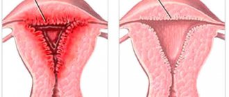
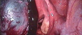
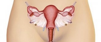
Mucinous cystadenoma is a benign ovarian tumor. Macroscopically, it is represented by a multi-chamber structure filled with gel-like mucus. This feature is due to the fact that the inside of the tumor chamber is lined with epithelium, morphologically similar to the epithelium of the cervical canal, which produces vaginal mucus.
- Symptoms
- Why is mucinous cystadenoma dangerous?
- Causes of development of mucinous cystadenoma
- Diagnosis of mucinous cystadenoma
- Treatment
- Consequences and prognosis
Mucinous cystadenoma often affects both ovaries. It can reach gigantic sizes, up to 40-50 cm. The neoplasm can occur in patients of any age, but more often it affects women after menopause.
What is cystadenoma
Many people think that this neoplasm is an ordinary benign ovarian cyst, but the danger of such a cyst is that it can malignize, that is, degenerate into a malignant neoplasm. Initially, the tumor is a bubble that has the structure of a cyst. That is, it is a capsule filled with liquid. This ovarian pathology can affect women of childbearing age, as well as those in menopause. Every woman should know what causes, symptoms, and how cystadenoma is diagnosed and treated.
What is characteristic of endometrial cancer?
According to statistics, 10% of endometrial cancer cases are mucinous type adenocarcinoma. It usually appears at the first stage of its development, which allows for a timely response to the appearance of a pathological formation. Women face this disease because the cells of the internal mucous membrane of the body of the reproductive organ undergo abnormal changes. Consultation with an Israeli specialist What is special about mucinous endometrial adenocarcinoma? In the course of histopathological studies, it is possible to establish that the presence of a dominant component of cells belonging to the endocervical type is characteristic. There are cases when mucinous metaplasia turns out to be benign. As for the tumor itself, it is assessed as highly or moderately differentiated. Its structure is complex, glandular, ciliary, and sieve-like. Characteristic are cystic expansions that are filled with mucus produced by the gland.
The main symptoms that women with a similar diagnosis have to face are frequent aching pain in the lower back, heavy menstruation for a long time. The described diagnosis can be made both to females of childbearing age and to those who are experiencing postmenopause. Medical practice shows that the disease is treated quite successfully, especially if you seek help in a timely manner.
From this we can conclude that if you feel the slightest discomfort, you should immediately consult a doctor. He has the necessary knowledge and skills to recognize cancer and refer it for laboratory and instrumental diagnostics. This way, it will be possible to detect the tumor at an early stage, and most importantly, get rid of it with little loss.
Reasons for development
The exact reasons for the development of this pathology are unknown, but the most likely occurrence of ovarian cystadenoma is during menopause, because the ovaries during menopause produce less hormones that prevent the occurrence of cancer.
Other factors that provoke the development of cystadenoma include:
- early puberty, in particular the onset of menstruation,
- late onset of menopause,
- infertility,
- frequent inflammatory and infectious diseases of the genitourinary system,
- abortions and miscarriages,
- constant heavy physical and psychological stress,
- exposure to carcinogens (including smoking),
- uncontrolled use of hormonal drugs, including contraceptives.
It is impossible to say for sure which woman will develop cystadenoma and which will not. Often it all depends on a combination of factors. Heredity plays a role. Women who have a family history of any type of ovarian tumor are more susceptible to cystadenoma.
Prevention of pathology
Prevention of the occurrence of pathology comes down to minimizing the impact of provoking factors. A woman should lead a healthy lifestyle, not overexert herself emotionally or physically, be faithful to her partner, thereby eliminating the occurrence of various infectious diseases of the genital area, and avoid unwanted pregnancy by protecting against it (abortion prevention). And, of course, you must not neglect visits to your gynecologist. This should be done at least twice a year.
Classification
Dangerous lesions are more often localized in the right appendage, since it has a better blood supply. Cystadenoma of the left ovary is extremely rare, mainly in cases of a malignant process that affects both appendages, which may indicate a malignant course of the pathology.
In the ovaries there may be functional cysts that are released from the follicles; cystadenoma differs from them in that it begins to develop from the epithelial tissue of the appendage. The classification of cystadenoma is determined by the nature of formation, structure and internal contents:
- Borderline ovarian cystadenoma is a transition between benign and malignant disease. In most cases, such tumors relapse into malignant ones after treatment, even if they were benign before therapy.
- Rough papillary cystadenoma is a morphological variety of the serous type of tumor, which has a whitish papillary vegetation. Thanks to these papillae, doctors can determine what stage of development the disease is at and select effective treatment.
- A pseudomucinous cyst is a type of mucinous cyst, but the difference between a pseudomucinous tumor and a mucinous one is that inside it there is pseudomucin - a mucoid substance, and not real mucin.
Depending on the type of cystadenoma and complications of the ovarian cyst, treatment tactics are selected.
Mucinous cystadenoma
These ovarian tumors have a multi-chamber structure and can reach sizes from forty to fifty centimeters. In this case, mucinous cystadenoma of the ovary can weigh ten or fifteen kilograms. Inside the capsule there is a large amount of mucin - a thick viscous secretion. Such a neoplasm is borderline mucinous carcinoma because it often becomes malignant.
Serous cystadenoma
In another way, the serous form of the cyst is called simple or smooth-walled; doctors often call it a ciliepithelial cyst. It has a dense single-chamber capsule filled with serous fluid and is localized only in one appendage. The size of the cyst varies from four to fifteen centimeters. Such a cyst can occur at any age, but more frequent cases occur when it develops in women after fifty years of age.
Papillary serous cystadenoma
The difference between papillary cystadenoma and simple serous cystadenoma is the presence of papillae on the inner surface. The tumor itself measures from three and a half to twelve centimeters, there are papillae outside it, and growths in the cavity. This type of neoplasm is characterized by a high tendency to malignant cell degeneration.
General information
This disease is divided into several types:
- Moderately differentiated adenocarcinoma is a malignant pathology of organs with an average degree of development of cancer cells. Cells with this form are not capable of rapid division, so cancer is amenable to conservative and surgical treatment.
- Well-differentiated adenocarcinoma is considered one of the simplest malignant tumors. It develops slowly, so pathology may be suspected in the later stages. The structure of cells of highly differentiated carcinoma has some similarity with healthy cells of the affected organ. A tumor can be distinguished from a healthy cell by the elongated shape of the nucleus. With this type of tumor, the presence of metastases in other organs is only 2-4 percent.
- Low-grade adenocarcinoma is a malignant neoplasm that develops in the epithelial tissue of the gland. A low degree of differentiation causes aggressive and rapid growth of the tumor. Papillary adenocarcinoma can be recognized when taking material for analysis. Tumor cells look like papillae of different shapes. Papillary adenocarcinoma forms in any internal organ; the peculiarity of such a tumor is its diversity of structure.
Microscopic studies of adenocarcinoma gave impetus to the development of oncology and the identification of different types of tumors. It is obvious that tumors have different structures, and cells multiply and progress differently. Cells and tissues of neoplasia gave the basis for classification of formations, in which a special place was occupied by malignant neoplasms of the glandular epithelium - adenocarcinoma, a frequently occurring type of cancerous tumor formation. The cells are mainly localized in the lymphatic vessels.
Symptoms
At the very beginning of the development of the pathology, the woman does not feel any discomfort in the area where the cystadenoma is located. As the cystic formation increases and damages the ovarian tissue, pain begins to appear in the lower abdomen, legs and lumbar spine. A neoplasm that is large in size leads to compression of nearby organs; the cyst causes the feeling of a foreign object in the abdominal cavity.
The woman's abdominal circumference increases greatly, since papillary cystadenoma is characterized by the development of ascites (the release of fluid into the abdominal cavity). A neoplasm of any type has a number of common symptoms:
- pain during urination and defecation,
- frequent urination,
- stool disorders - constipation or diarrhea,
- menstrual cycle irregularities,
- bloody discharge between periods,
- feeling of tightness in the lower abdomen.
The pain syndrome is localized on the side of the affected ovary, but can radiate to the lower back, pubis, sacrum, and groin area. If a cyst ruptures or its leg becomes twisted, the pain becomes severe and the body temperature rises. In this condition, the woman should be immediately hospitalized and operated on.
Types of tumors and their features
There are several types of cystadenomas, differing in structure and composition, as well as in the symptoms they exhibit.
Serous
There are two types of serous cystadenoma: simple serous smooth-walled and papillary.
Simple smooth wall
Serous ovarian cystoma (also called smooth-walled celioepithelial cystadenoma or serous cyst) is considered the most harmless neoplasm. It is a true benign ovarian tumor. It is formed from previously unresolved follicular or luteal cysts, which normally should regress within several months (on average, over three to four cycles). Previously, this type of cyst was found in women over thirty years of age, but now the disease has become “younger” and is detected even in twenty-year-old girls.
The surface of the tumor is smooth, and its serous contents are in most cases transparent and yellow in color. Most often it has one chamber and affects one ovary. It is quite mobile and does not cause pain. It can be located to the left or right of the body of the uterus (depending on which ovary is affected) or is found behind it. As a rule, small serous cystadenomas do not bother women. As the size of the tumor increases, general symptoms gradually appear.
Serous cystadenoma has a smooth surface and one chamber
Papillary - papillary and rough papillary
Papillary papillary and papillary rough papillary cystadenomas are a type of serous type cystadenomas. But they stand out separately. Such neoplasms are usually multi-chambered and on the surface lining the cavity from the inside there are quite dense papillary (papillary) outgrowths of a whitish hue on a wide stalk. These growing seasons can be either single or numerous. A feature of coarse papillary growths is the absence of the likelihood of tumor degeneration into cancer.
Papillary papillary cystadenoma, on the contrary, is capable of malignancy (malignancy). This is due to the fact that the outgrowths have a soft consistency and often merge with each other, forming peculiar tumors (nodes) that can grow outward through the wall of the capsule, resembling cauliflower in appearance. The spread of the tumor to neighboring organs indicates malignant degeneration. As a rule, such cystadenomas form on two ovaries. Initially, the tumor is mobile. As its size increases, mobility becomes limited.
On the ultrasound image, the doctor will clearly see papillary growths
Mucinous
Mucinous cystadenoma (or pseudomucinous tumor) refers to benign epithelial neoplasms of the ovary. Most often detected during postmenopause. The tumor is usually multi-chambered, filled from the inside with mucinous contents, which is a mucous or jelly-like substance of a yellow or brown (due to blood content) hue with the inclusion of special proteins - glycoproteins and heteroglycans. The surface is even and smooth outside and inside. Usually reaches impressive sizes; there are cases when the formation grew to thirty centimeters in diameter or more. At the same time, its wall was thin and transparent.
Additionally, borderline mucinous tumor, pseudomyxoma of the ovary and peritoneum, and Brenner's tumor are distinguished, which have a high risk of degenerating into cancer.
Mucinous cystadenoma has a capsule of uneven thickness and many chambers
Borderline mucinous cystadenoma
Borderline ovarian cystadenoma is potentially malignant. It has a smooth surface outside and inside, with a multi-chamber structure. The cells of the inner membrane are capable of proliferative growth. In this regard, the risk of malignant degeneration of the tumor increases. As a rule, such cystadenomas are not characterized by invasive growth, that is, they do not grow into surrounding organs and tissues.
Pseudomyxoma of the ovary and peritoneum
Pseudomyxoma of the ovary and peritoneum is considered a fairly rare tumor, usually affecting women over fifty years of age. The main feature of the neoplasm is that it is not capable of infiltration and germination into surrounding organs and tissues, so it is difficult to talk about the malignant nature of such a cystadenoma.
Recent Entries
Is it possible to give a mirror: how to protect yourself from bad omens It became known about the influence of cell phone towers on human health Is it possible to eat bananas bought in Russia?
Pseudomyxoma has a thin capsule that can rupture spontaneously or during a bimanual examination by a gynecologist. In this case, pseudomucin - the jelly-like contents of the tumor chambers - enters the abdominal cavity, settling on organs and tissues. Its particles become overgrown with capsules, blood vessels and nerves. In this way, new pseudomyxomas are formed.
Brenner's tumor
Brenner's tumor is also called fibroepithelioma or mucoid fibroepithelioma. The tumor is formed from the ovarian stroma - connective tissue containing blood vessels. The peculiarity of this neoplasm is that it can appear in the fair sex at any age: at fifty or five. It can reach large sizes and most often appears on the left ovary. It has an ovoid (round or oval) shape and a smooth shiny surface. As a rule, it is rare and characterized by a benign course. But the possibility of malignant degeneration cannot be ruled out.
The tumor is named after Franz Brenner, who first described it.
Malignant cystadenocarcinoma
Malignant cystadenocarcinoma is a malignant tumor resulting from the degeneration of serous or mucinous cystadenoma.
Serous cystadenocarcinoma is the most common and is detected in 70% of cases, characterized by accelerated growth and rapid spread to surrounding tissues and organs. Mucinous cancer is detected much less frequently (in approximately 10% of cases) and is characterized by slow development and asymptomatic course in the initial stages. When large in size, it causes discomfort in the lower abdomen, similar to those that occur with intestinal problems.
Diagnostics
To make a diagnosis:
- gynecological examination,
- ultrasonography,
- computed or magnetic resonance imaging,
- blood test for tumor marker CA-125.
After an accurate diagnosis has been made, the doctor can prescribe treatment and choose the type of operation that will be performed on the patient.
Stages
Malignant tumors are divided into 4 stages of development:
- The first degree of adenocarcinoma is characterized by the location of the tumor exclusively within the ovarian tissue and glandular capsule. The neoplasm usually does not manifest itself in any way and becomes an accidental finding during a gynecological examination or ultrasound.
- At the second stage of development, the cancer increases in size and goes beyond the boundaries of the organ. Cancerous elements are found in the fallopian tubes, uterus, pelvic tissues, and neighboring organs.
- Stage 3 serous cystadenocarcinoma of the ovary is characterized by the spread of the pathological process beyond the pelvis to the sheets of the peritoneum or to the lymph nodes.
- Regardless of the size of the primary tumor, with stage 4 disease development, distant metastases are detected outside the abdominal cavity.
Bilateral ovarian cystadenocarcinomas do not affect the stage of the disease. When making a diagnosis, the spread of cancer cells beyond the ovarian glands is critical.
Treatment methods
For cystadenoma of the appendages, treatment can only be performed surgically. Without surgery, it is impossible to get rid of the tumor either with the help of drugs or traditional medicine. The operation involves surgical removal of the tumor, sometimes including the ovary. The type and extent of surgical intervention depends on the type of cystadenoma, its size, clinical picture and the degree of likelihood of malignancy.
Laparoscopy
Laparoscopy is justified for simple serous neoplasms of a benign course, the size of which does not exceed four centimeters. This type of operation helps preserve the ovary, and with it the reproductive function. This method of surgical intervention is minimally invasive, since the size of the incisions on the abdomen does not exceed one and a half centimeters. After laparoscopy, a woman can plan a pregnancy in a few months.
Laparotomy
This type of surgery is an abdominal operation in which one large incision is made in the abdominal wall. This treatment is carried out for large cystadenomas, with a risk of malignancy, as well as for patients during menopause. During the operation, the tumor, ovary, and uterine tubes are excised.
A neoplasm in women after fifty years of age is treated by removing two appendages, which prevents the occurrence of cancer on the second ovary. After this, the patient is prescribed hormonal therapy, which helps replace the natural hormones previously produced by the ovaries. Women of childbearing age after removal of one ovary are able to conceive, carry and give birth to a child.
Types and symptoms of cystadenocarcinoma
In modern medicine, there are two types of cystadenocarcinomas: serous and mucinous. A type of serous cystadenocarcinoma of the ovary, also called serous type ovarian cancer, occurs in more than sixty percent of patients with malignant tumors. In most cases, this disease develops as a result of the transformation of benign epithelial cells of a cystic formation into malignant ones. Mucinous tumor is quite rare. It is diagnosed in only fifteen percent of patients with malignant neoplasms in the pelvic organs.
Important! The symptoms of each type of ovarian cystadenocarcinoma depend on the stage of the disease. The sooner the patient detects them and consults a doctor, the more favorable the outcome will be.
Mucinous tumors are predominantly asymptomatic and are therefore detected in the later stages, when pain appears in the lower abdomen and ascites develops. Patients often complain of impaired intestinal function, accompanied by constipation or indigestion, and a feeling of constant discomfort in this part of the body. On ultrasound, a mucinous malignant neoplasm appears as a tumor with an uneven consistency. During palpation in this case, the patient will feel pain in the place where the mucinous body has formed. During a rectovaginal examination, the doctor can observe tumor nodes.
Treatment during pregnancy
A woman with cystadenoma has every chance of carrying a pregnancy to term and giving birth if the tumor is small. For tumors up to three centimeters in size, doctors monitor the tumor. If a cystadenoma begins to progress rapidly, it is urgently removed, since its growth can lead to displacement of the uterus, twisting of the pedicle and rupture of the tumor walls.
Laparoscopy is performed after the fourteenth week of pregnancy, when the placenta has already fully formed. Before this period, surgical intervention is carried out only according to strict indications (twisting of the leg, suppuration, rupture of the capsule) in order to save the woman’s life. Pregnancy in such cases is usually terminated.
Description
Mucinous cystadenoma has a somewhat similar structure to an ovarian cyst. A distinctive feature is multi-chamber, which is clearly visible during an ultrasound examination. The contents of each chamber are presented in the form of mucin - mucous contents. The tumor shell is prone to rapid growth, and therefore the tumor can reach sizes of 15-50 cm in diameter.
Mucinous cystadenoma of the ovary is a borderline type of tumor due to the risk of its degeneration into a malignant formation.
Consequences
The development of cystadenoma can lead to various complications and consequences, including:
- dysfunction of the appendages,
- infertility,
- malignancy of the process,
- disruption of the functioning of neighboring organs - the uterus, second ovary, intestines, bladder,
- inflammatory processes in the abdominal cavity,
- compression of blood vessels by the tumor,
- phlebeurysm,
- the formation of nodes and the formation of blood clots.
Sometimes tumors do not cause complications, but acute complications often occur that require emergency removal of the tumor:
- peritonitis of the abdominal cavity due to rupture of the walls of the neoplasm,
- necrosis of cystadenoma due to twisting of the leg,
- purulent inflammatory process in cystadenoma.
These conditions pose a direct threat to the patient’s life, so you should immediately call an ambulance if the following signs of acute complications occur:
- hyperthermia,
- acute pain,
- increased sweating,
- increased heart rate,
- feeling of fear,
- overexcitement followed by lethargy,
- nausea and vomiting,
- stool retention,
- darkening in the eyes,
- fainting,
- severe weakness
- chills,
- a sharp decrease in blood pressure.
If emergency measures are not taken for these complications, death occurs no later than within a couple of days.
Is it possible to treat ovarian cystadenoma without surgery?
Cystadenoma cannot be treated without surgery; its necessity is caused by:
- the risk of progression to cancer;
- growth and compression of the pelvic organs;
- difficulty in removal if complications develop (peritonitis, destruction, bleeding).
The most gentle and effective method of removal is laparoscopy, since:
- less surgical trauma;
- less often then adhesions form;
- short recovery period;
- lower risk of infection and postoperative hernia.
Laparoscopic surgery to remove an ovarian cyst
An endoscope is inserted into the abdominal cavity through small incisions to help definitively confirm the diagnosis. If possible, simultaneous dissection and removal of the tumor is performed. If there are contraindications, the intervention is converted to open - the anterior abdominal wall is dissected. This type of operation is indicated for:
- doubts about the benignity of the tumor;
- sizes from 12 cm;
- obesity;
- extensive adhesive process (occurs after inflammation, injury, surgical treatment).
The need for open access also arises when signs of malignancy are identified during urgent histology. In this case, complete removal of the ovary is necessary, and then chemotherapy, radiation, and hormonal therapy. Similar tactics are required when identifying papillary and mucinous cystomas, and after menopause, the uterus and second ovary are simultaneously removed.
Open surgery to remove an ovarian cyst
In young women, with reliable confirmation of benignity and serous cystadenoma, only the tumor itself is excised and the ovary is sutured, which does not subsequently interfere with pregnancy and childbirth.
Complications from formations
Complications requiring emergency abdominal surgery include:
- torsion of the tumor leg - sharp pain, tension in the abdominal wall, nausea, vomiting, loss of consciousness;
- rupture and bleeding - happens with rapid growth, injury, manifested by pain with a sudden drop in pressure, rare and weak pulse, pale skin.
Delay in these cases leads to inflammation of the peritoneum - peritonitis, and there is a risk of death if surgical treatment is refused. An unfavorable option also occurs when a mucinous tumor ruptures and cells spread throughout the peritoneum (pseudomyxoma).
In some cases, they acquire the ability to form fluid, causing constantly recurrent ascites, the need for frequent puncture and pumping.
Symptoms of the painful condition
At the initial stage of its development, the disease is asymptomatic or the clinical phenomena are weakly expressed, and only as the pathological process worsens do the first signs appear.
However, they have no characteristic differences from the symptoms of ordinary breast cancer:
- Chest pain.
- Nausea.
- Fast fatiguability.
- Weight loss.
- Nipple discharge.
- Minor swelling.
- Deformation of the patient's breast.
- Erosion, peeling and crusts on the nipples.
- Inflammation of the lymph nodes.
- When palpating the gland, uncharacteristic nodules are felt.
- Redness of the skin over the tumor site.
The only exception is the good mobility and soft structure of the neoplasm.
Distinctive features of the malignant condition
Among other clinical features of cancer, it is necessary to note:
| Main settings | Character |
| Development rate | Slow. |
| Infiltration in the axillary lymph nodes | Absent. |
| Outlines | Clear rounded. |
| Skin connection | Absent. |
| Type of education | Resembles a knot. |
| Degree of early malignancy | Low. |
| Modification of breast skin | Not visible. |
| Internal Contents | Soft and mobile consistency. |
| Tumor size (diameter) | From 1 to 20 cm. |
| When cutting the seal | A shiny mucous mass flows out. |
A scientific study of the tumor body showed that estrogen receptors are present in 60% of the cells. In other words, the neoplasm is hormone-dependent, that is, its active growth is provoked by this hormone.
However, individual cells can form:
- Heavy.
- Threads.
- Papillae.
- Alveoli.
Specific signs of cystadenocarcinoma
Late-stage ovarian cancer is accompanied by the following specific symptoms:
- Disrupted menstrual cycle, in which uterine bleeding becomes either too heavy or, conversely, rare.
- Malfunctions of the bladder and intestines.
- Increased abdominal volume for no reason.
- Periodic increase in body temperature, especially in the evening.
- Loss of body weight due to disruption of the gastrointestinal tract.
- Deterioration in well-being, which manifests itself in rapid fatigue, constant fatigue, apathetic states, and drowsiness.
We recommend you learn: What is ovarian cystadenoma


