The development of the incredibly complex human body begins with a single cell. The beginning of a new life is laid at the moment of fusion of two small gametes - male and female, sperm and egg. However, for fertilization to occur, germ cells must go through a long path of formation and maturation.
The egg, the female gamete, is laid and grows in the ovary, a specialized organ of the female reproductive system. There it is surrounded by an island of cells that form the ovarian follicle. The cell lives in the ovary, surrounded by follicular fluid and membrane until ovulation.
Embryonic oogenesis
The development of female germ cells (ovogenesis, oogenesis) begins at the embryonic stage of the formation of the body of the future woman. At this time, the eggs are not yet as large and mature as they become at the time of possible fertilization. Now these are only the rudiments of future eggs - oogonia (or oogonia). They are located in the deep layers of the ovary and reproduce through mitosis.
Oogonia are ordinary cells that have a normal double set of chromosomes, like the rest of the body cells. Before they can give birth to a new person, they will have to lose half of this set.
When a girl is born, her ovaries already contain hundreds of thousands of primary oocytes, most of which will die before they even have time to develop. How many eggs will survive to full maturity? Only 400–500 cells.
Where and how does it form in humans?
The question of how many eggs a woman has is of no practical importance. Because their number is not so important as how many of them mature and can become the beginning of a new life.
Their formation and maturation occurs in a woman’s ovaries. Even in the prenatal period, a girl develops about a million of these cells. The older the girl gets, the more reproductive gametes die. By the time of puberty, there are about 400 thousand of them.
During the entire reproductive period of a person, they continue to die and no more than 300 eggs enter the maturation process. Therefore, the number of children a woman can conceive is limited.
Development of eggs in the body of a mature woman
After the birth of a girl, new eggs are no longer laid (unlike sperm, which are actively produced in men throughout their lives). How many cells are formed in the embryo, so many will take part in the reproductive function.
Until the onset of puberty, a woman lives without serious changes in the ovaries, the primary oocytes quietly wait in the wings. And then the first egg matures and the cycle begins.
The female menstrual cycle is a special form of organizing the maturation of germ cells. Once it is fully formed, the period between its beginning and end will take approximately 28 days. The duration may vary depending on the individual characteristics of the body, but normally it is about a month, which is why the cycle is also called monthly.
Every month, several follicles with small eggs sleeping inside them are activated and begin to develop. Exactly how much growth will take place is an individual indicator for each woman. First-order oocytes divide in a special way (meiosis) and lose half of their chromosome set, becoming haploid, that is, containing a single set of chromosomes.
How many chromosomes does a person have? There are 46 of them in each cell of the body, connected in pairs. To maintain genome constancy, gametes have only half of this number - 23 chromosomes. Merging, they form a full-fledged zygote.
The primary follicle is then called secondary; fluid accumulates in its cavity, forming a vesicle.
How many stages does the future egg go through in its development to the point of being fertilized? These are stages such as
- oogony, that is, a germ cell formed at an early stage of embryonic development;
- first-order oocyte, when the germ cells have completed mitotic divisions and accumulated the necessary nutrients;
- a second-order oocyte with half the set of chromosomes;
- mature oocyte.
During one cycle, follicles are normally activated in one ovary (left or right). Afterwards they alternate.
Of several active follicles, only 1–2 reach the mature vesicular stage, the rest undergo reverse development. A large vesicle of a mature follicle is visible on the surface of the ovary as a rounded bulge about a centimeter in diameter. The egg in it is almost mature, increased in volume due to accumulated nutrients (yolk), and almost ready for fertilization. There are only a few hours left before its release.
Structure and functions of the egg.
Home Favorites Random article Educational New additions Feedback FAQ⇐ PreviousPage 14 of 17Next ⇒
The eggs are immobile because have a spherical or slightly elongated shape; consists of a nucleus, cytoplasm, several membranes; the volume of the nucleus is less than the volume of the cytoplasm; much larger than a somatic cell. Discovered by K. Behr in 1827.
The outside is covered with several shells:
- external - follicular, formed by follicular epithelial cells,
- internal - shiny, so called due to its optical properties. The outgrowths of follicular cells penetrating the membrane are in contact with the cytoplasm.
- in the center of the egg there is a nucleus with nucleoli, surrounded by cytoplasm. The cytoplasm contains a huge number of inclusions in the form of yolk granules, as a supply of nutrients necessary for the development of the embryo.
Shell functions:
- Protective;
- Provides the necessary level of metabolism;
- Prevents the penetration of more than one sperm;
- Promotes the introduction (implantation) of the embryo into the wall of the uterus;
- Maintains core shape.
Size: in some animals the eggs are so large that they are visible to the naked eye - eggs of fish, amphibians, eggs of birds and reptiles.
In modern animals - the herring shark has an egg up to 30 cm and the ostrich.
Small size - in animals in which, during the development of the embryo, it receives nutrients from the mother's body - higher placental animals.
Shells by origin: primary, secondary, tertiary.
1. Primary - a derivative of the cytoplasm - vitelline, characteristic of all animals.
2. Secondary - due to the activity of cells that feed the egg - the chitinous shells of arthropod eggs.
3. Tertiary - arises as a result of the activity of the female gonads: shell, subshell - the protein shell in the eggs of birds and reptiles, as well as the gelatinous shell in amphibians.
The structure of a bird's egg.
An egg is divided into yolk, white, shell membranes and shell. The yolk makes up 32-36% of the total mass of the egg. There is a pronounced layering in the structure of the yolk. In the center of the yolk there is a pale yolk ball - latebra, consisting of a light yolk, which is connected by a cord to the embryo - blastoderm, located on the surface of the yolk in the form of a white opaque formation. The latebra is successively surrounded by alternating layers of light and yellow yolk. The number of such layers is about 6. The peripheral part of the yolk is covered with a vitelline membrane, separating the yolk from the white. All the most biologically valuable substances are concentrated in the yolk. It contains all the lipids of the egg, all the essential vitamins and microelements. The shell consists of a mixture of calcium carbonate salts and calcium and magnesium phosphate salts.
Types of eggs:
Depending on the amount of yolk and the nature of its distribution in the cytoplasm, several types are distinguished:
- Isolecithal - a small amount of yolk is evenly distributed throughout the cytoplasm: primary isolecithal (for the egg of echinoderms, mollusks), secondary isolecithal (mammalian cells).
- Alecithal - without yolk. They feed on follicular cells, which is typical for mammals.
- Telolecithal - the bulk of the yolk accumulates in the lower vegetative pole, while the nucleus and cytoplasm are in the upper animal zone, without yolk. Moderatetelolecithal (the yolk is immersed in the cytoplasm, and not separated from it in the form of a separate mass - sturgeons, amphibians). Absolutely telolecithal (the yolk is completely separated from the cytoplasm, the egg of birds, and oviparous mammals)
- Centrolecithal - have the yolk concentrated in the center of the cell, while the cytoplasm forms a thin surface layer, amphibians.
Fertilization.
Fertilization is the process of fusion of female and male reproductive cells, resulting in the formation of a fertilized egg - a zygote.
Consequences of fertilization:
- Activation of the egg is an incentive for development.
- Synkaryogamy is the formation of a diploid nucleus as a result of the fusion of the haploid nuclei of germ cells carrying the genetic information of both parents.
In order for fertilization to occur, sperm need to meet an egg. Insemination is the process of meeting female and male reproductive cells.
Insemination:
· External - meeting of sperm and egg in the external environment (amphibians, fish).
· Internal - sperm are introduced by the male into the female’s reproductive tract during sexual intercourse (land dwellers, higher vertebrates, reptiles, mammals).
· Mixed - the sperm is enclosed in a special “package” - a spermatophore, it enters the water, then is swallowed by the female’s cloaca, as a result of which the sperm end up inside the female’s body (caudate cephalopods).
Fertilization in animals.
- The rapprochement phase: it is based on the release of special chemical substances by the sex cells - gamones - which activates their rapprochement, the production of gamones is determined by the selectivity of fertilization of the egg by a sperm of the same species. Eggs secrete gynogamones, and sperm produce androgamones. These substances act as an antigen-antibody system according to the principle of complementarity. Some of these substances are activated by the movements of sperm, others promote their fusion and also dissolve their nuclear membrane.
- Penetration phase: begins with the penetration of one (monospermy) or several (polyspermy) sperm into the egg. There are 2 methods of penetration:
- 1 method is less common: through micropiles (penetrating holes in the membrane of the egg through which sperm penetrate), only for some species.
- Method 2 - for most species - penetration of sperm as a result of the acrosomal reaction, which occurs as a result of contact of the head of the sperm with the surface of the egg.
Upon contact with the egg, the acrosome shell is destroyed, the acrosomal filament is released from it, and enzymes are released that destroy the egg shell. The acrosomal filament penetrates the dissolved membrane and fuses with the membrane of the egg. At this point, an ovoid tubercle is formed from the cytoplasm of the egg. It captures the nucleus, centrioles, and mitochondria of the sperm and draws them into the egg. After the formation of the so-called fertilized cell membrane, other sperm can no longer penetrate the egg. The sperm nucleus swells and acquires the features of an interphase nucleus. This nucleus is called the male pronucleus.
The phases of internal fertilization can be divided into 2 stages:
- The stage of two pronuclei - morphologically separate male and female eggs. By this time, the egg has not yet completed the 2nd meiotic division, so its viscosity increases, which prevents the rapid convergence of the nuclei.
- Synkaryon - the male and female nuclei closely fit together - the nuclei merge, a diploid synkaryon (zygote) is formed. After the formation of a synkaryon, the zygote begins fragmentation (mitosis) and the embryonic period of development of organisms begins - embryogenesis.
Double fertilization of plants and development of germ cells.
The process of formation of germ cells in plants consists of two stages:
- Sporogenesis.
- Gametogenesis.
Fertilization in flowering plants is represented by the formation of male and female gametophytes.
The main stages of double fertilization:
- Formation of male and female gametophytes.
- Pollination.
- Double fertilization and seed formation.
Gametophyte formation.
A) male gametophyte.
1. Microsporogenesis occurs in the anthers of stamens. The stamen is a microsporophyll, consisting of a filament, at the top of which there is an anther - microsporangium - an organelle where microspores are formed.
Consists of 2 parts. In microsporangia in the mother cell (2n), as a result of meiosis, 4 haploid microspores are formed from one diploid cell. After meiosis, microspores are covered with two membranes, the outer one is eczyma, the inner one is entyma.
2. Megagametogenesis consists of two successive mitotic divisions of the microspore nucleus; as a result of the first mitotic division, two cells are formed: vegetative and generative. Only the generative cell undergoes the second mitotic division, from which 2 sperm are formed. In this way, a male gametophyte is formed - a stigma grain, consisting of 3 haploid cells (1 vegetative and 2 sperm).
B) formation of the female gametophyte
1) Megasporogenesis occurs in the ovary of the pistil. On the inner surface of the pistil ovary there are ovules-megasporangia, in the central part of which a megaspore is formed.
Structure of the ovule
It consists of: outer integuments (intiguments) above the upper pole of the central part there is a pollen opening (micropyle) and a nucellus (central part), in which the female gametophyte of the megasporangium is formed and germinates and where the embryo is formed, it communicates with the wall of the ovary using an achene. In the tissues of the ovule, the mother cell of the macrospore (2n) is isolated, from which 4 haploid megaspores are formed by meiosis, one of which continues further development and leads to the formation of a female gametophyte, and three die.
2) Megagametogenesis.
The initial cell (megaspore) undergoes 3 mitotic divisions, and karyokinesis is not accompanied by cytokinesis; as a result of 3 divisions, 1 cell is formed containing 8 haploid nuclei, that is, a female gametophyte (embryo sac) is formed. At each pole there remain 3 nuclei, and 2 nuclei remain in the center, then they merge to form a diploid nucleus, a cell membrane is formed around each nucleus, thus the female gametophyte consists of 8 cells: 3 cells of the egg apparatus (including 1 egg(nc ) and 2 accompanying synergids). The egg apparatus is located at the upper pole of the gametophyte; at the other pole there are 3 antipodal cells (nc), which do not participate in fertilization and later die. In the center is a diploid cell.
Pollination.
Precedes fertilization. Pollination is the transfer of pollen from the anthers of the stamens to the stigma of the pistil; there is cross-pollination and self-pollination.
Crossing is the transfer of pollen from the anthers of the stamens of one plant to the stigma of another plant, with the help of insects, wind, and water. It is found more often in nature and provides genetic diversity in the offspring.
Flower adaptation: bright color, smell.
Self-pollination is the transfer of pollen from the anthers of the stamens to the stigma of one flower. Rarely seen.
Fertilization.
Fertilization precedes the germination of pollen grains and the formation of a pollen tube. Germination begins with swelling of the grain and the formation of vegetative cells. The long extension of the pollen tube reaches the embryo sac. Two sperm cells, formed during the division of the generative cell (pollen grain), move along the pollen tube channel. The tip of the pollen tube secretes enzymes that soften the tissues of the stigma and style; ultimately, the pollen tube penetrates through the micropyle into the embryo sac, and the membranes dissolve. The sperm fuses with the egg to form a zygote, which gives rise to the embryo of the seed. The second sperm fuses with the diploid central cell to form triploid tissue, giving rise to the endosperm.
The essence of double fertilization: the first sperm fuses with the egg, forming a zygote, from which the seed embryo further develops; the second sperm fuses with the central cell to form (3n) tissue, giving rise to the endosperm.
This process is called double fertilization - fertilization in flowering plants, in which the egg is fertilized by one sperm, and the diploid nucleus of the central cell by another sperm. The process was discovered in 1898 by S.G. Novashin.
Biological significance:
1. Significant savings in energy resources; thanks to the triploid endosperm, the ovule does not store nutrients for future use.
2. Faster growth of polyploid tissue and accelerated development of the embryo.
A fertilized egg is a seed; the wall of the ovary is the pericarp; ovary - fruit. Number of seeds = number of ovules.
Forms of reproduction.
Reproduction is the ability of living organisms to reproduce their own kind to ensure continuity and continuity of life.
Forms.
The classification is based on the type of cell division (mitosis - asexual, meiosis - sexual)
- Asexual reproduction is various forms and methods of reproduction of organisms, characterized by the absence of the sexual process and without the participation of germ cells.
- Sexual reproduction refers to various methods of reproduction in which an organism usually develops from a zygote formed by the fusion of sex cells.
Table No. 11. Comparative characteristics of asexual and sexual reproduction.
⇐ Previous14Next ⇒
Preparing the body for pregnancy
While the egg is diligently maturing in the ovary, the female body is already planning a future pregnancy. To provide the one-celled embryo with a soft landing, it builds up an additional layer of mucous membrane in the uterus, abundantly supplying it with blood vessels.
If two germ cells meet in the sacred act of fertilization, a zygote will be implanted into this layer. If conception does not occur, the body will have to get rid of excess cells after the end of the cycle. When they are separated, blood vessels rupture. This is menstrual bleeding.
You should remember when conception is possible
Conception is possible if it is wet and there is mucus. Conception is impossible if it is dry.
This dryness appears twice: the first time - after the end of menstruation and then does not last long; the second time - 3-4 days after ovulation and lasts until the end of the cycle, i.e. before the start of a new menstruation.
Cervical position
The next sign of fertility is a change in the position of the cervix.
The position of the cervix can be determined not only by a doctor. Any woman, using specific advice from a specialist, can learn to assess the position and consistency of the cervix, as well as the degree of its openness. Lots of women do this very well. This is especially useful for those who are breastfeeding, are close to menopause, and are also experiencing difficulty getting pregnant.
Before the period of ovulation, the cervix is dry, hard, lowered towards the bottom of the vagina, the external cervical opening is closed. All these are characteristic signs of an infertile period.
During the period of ovulation, the cervix becomes soft and moist (due to the abundance of mucus), the opening of the cervical canal expands, and it itself rises upward. Immediately after ovulation, under the influence of the hormone progesterone, the cervix very quickly becomes hard again, goes down, and its external opening closes. With some skills, all these changes can be easily observed on your own.
Remember: the cervix can be as hard as the tip of your nose during the infertile period, and as soft as lips during the fertile period.
What to read?
Release of a mature egg from the ovary
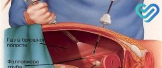
The vesicular follicle enlarges to its maximum, raising the integumentary epithelium of the ovary. Now it is called the Graafian vesicle and contains a completely mature reproductive cell. At a certain point, the membranes rupture and the egg is released into the body cavity.
This critical moment of release is called ovulation of the egg. A mature oocyte leaves the follicle, which has kept it from external influences for so long, and begins an independent life.
But what should an egg do in a body cavity? How can she meet the sperm? Nature, of course, took care of this: the egg lives in the abdominal cavity for only a few minutes, after which it is drawn in by the funnel of the fallopian tube, equipped with a fringed border.
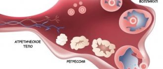
The burst follicle does not disappear from the scene; it still has an important role to play. In its place, the so-called corpus luteum is formed. This is a temporary endocrine gland that will supervise conception and slow down the development of other follicles by secreting the pregnancy hormone progesterone. If fertilization does not occur, the corpus luteum will collapse after a few days. If pregnancy has occurred, this gland will work for as long as it takes to form a full-fledged placenta, which will then take over its functions.
Fertilization of an egg is a process from A to Z in time
That is why the remaining trace on the ovary after regression of the temporary endocrine organ is called the white body.
What will happen during pregnancy
If the mystery of the fusion of male and female reproductive cells has occurred, then the corpus luteum will provide the following important functions:
- increasing synthesis of progesterone, ensuring the preparation of the uterus for the arrival of the embryo;
- suppression of the ovulatory ability of the ovary;
- production of hormones and biologically active substances necessary for the first weeks of the embryo’s life;
- fetal protection;
- maintaining pregnancy.
If the temporary endocrine organ works normally and produces a sufficient amount of necessary hormones, then this is quite enough for the pregnancy to develop calmly all the time until a full-fledged placenta is formed.
Typically, the highest hormonal activity of the corpus luteum occurs between implantation and 12 weeks of pregnancy. After this, the developing placenta gradually takes over all endocrine functions to ensure the growth and development of the fetus.
If the functioning of the temporary hormonal organ that produces progesterone is disrupted, it is necessary to start taking Utrozhestan or Duphaston in time. Thanks to any of these drugs, it is possible to maintain pregnancy with luteal insufficiency
It does not matter in which ovary ovulation occurred if the result is the formation of a full-fledged corpus luteum. The main thing is that this process becomes the main factor for the occurrence and maintenance of pregnancy
The temporary endocrine organ of the ovary, formed immediately after ovulation, creates maximum conditions for the onset of conception, ensuring the first days of the embryo’s life, implantation and development of the embryo.
In case of hormonal deficiency due to decreased function of the corpus luteum, the only real chance for preserving the life of the embryo will be the timely use of a drug that compensates for the lack of progesterone.
https://youtube.com/watch?v=MQLAaSXyOi4
Each ovary has hundreds of thousands of follicles. And, of course, each of these follicles is exposed to the same blood supply.
So your question...
Why is ovulation not seen in both ovaries together?
… such as:
Why is ovulation not seen in two follicles (independent of the ovary)?
All follicles are stimulated by FSH and they actually begin to mature at the same time. However, in a given cycle, usually only one follicle grows completely. Here we have competition between follicles.
Guyton is not my favorite physiology book, but this suggests an explanation for it:
only one follicle matures completely every month, and the rest undergo atresia:
After a week or more of growth—but before ovulation—one of the follicles begins to outgrow all the others; the remaining developing follicles involute (a process called atresia), and these follicles are said to become atretic.
How long does an egg live?
The most important issue that concerns all women planning a pregnancy or wanting to protect themselves from it. After all, fertilization is normally possible only for a certain period of time, when the egg makes its journey through the fallopian tube to the uterus. How long does this period last?
A mature female gamete is a fairly large round cell, reaching 150 microns in diameter. She has a significant supply of protein-lipid nutrients in the cytoplasm, thanks to which she lives until fusion with sperm and implantation into the uterine wall. The cell has enough yolk for 12–24 hours (sometimes a little longer) from the moment it leaves the follicle.
Thus, the egg lives after ovulation for about another day. How long can a sperm survive in the female genital tract? On average 3–4 days (its lifespan may vary).
For conception to take place, the sperm must already be waiting for the female cell in the uterus or hurry towards it.
Maturation of the egg after menstruation by day
To determine how many days after menstruation ovulation occurs, it takes a long time to observe the body. Each stage of egg growth is accompanied by certain signs. A few days after the end of menstruation, characteristic tingling sensations appear in the appendages. As the follicles grow, the pain may intensify. Closer to ovulation, the following symptoms are observed:
- Mucous discharge from the vagina;
- Increased sex drive;
- Mood changes;
- Heaviness in the chest;
- Increase in body temperature.
Immediately after the rupture of the follicular walls, the oocyte moves towards the uterus through the fallopian tubes. If it meets a sperm, a zygote is formed. Every day of its movement is accompanied by the process of cell division. When it enters the uterine cavity, an embryo sac is formed in it. The next step is to implant the cell into the uterus. This occurs 7-10 days after ovulation.
The maturation of the egg varies from day to day as follows:
- On days 5–7 from the beginning of menstruation, the cell is 4–5 mm in diameter.
- On days 8–10, a cover of connective tissue appears. The dominant stands out among other formations.
- On days 11–13, the diameter of the dominant reaches 16–18 mm. The remaining follicles regress and gradually dissolve.
- On days 14–16, the next ovulatory cycle begins. The diameter of the remaining follicular cell, called the Graafian vesicle at this stage, is 20–22 mm.
Not in all cases the female body works like a clock. The absence of ovulation during regular periods is called anovulation. If this phenomenon occurs no more than 2 times a year, there is no need to worry. Constant anovulatory cycles indicate disturbances in the body
Therefore, it is important to determine why there is no ovulation during regular periods. To do this, an extensive examination of the body is carried out
Size
The size of the egg at ovulation is three times the size of the sperm. According to scientists, the size is 0.1-0.15 microns.
YAC contains nutrients necessary for the development of the fetus in the first weeks of life. She also has a set of chromosomes. The ovary of a newborn girl contains up to 1.5 million follicles, some of which die before puberty. Until now, this relationship is not clear to scientists.
When the first menstruation occurs, no more than 400 thousand oocytes remain in the woman’s body. They mature throughout their lives. By the time the nuclear center is released, the size of the water capsule is 2 centimeters.
What does donor egg mean?
A donor egg is genetic material provided by a donor, a woman who voluntarily agrees to donate her egg to a woman suffering from infertility. The donor must meet all the parameters specified in the agreement between the clinic and the donor candidate.
Subsequently, specialists in the field of reproductive health will work with the obtained genetic material; it can be used in almost all modern assisted reproductive technologies.
The donor evaluation criteria are as follows:
- age coinciding with the period of active reproductive functioning (20-30 years)
- normal indicators of health as a system as a whole
- the woman does not smoke or drink alcohol
- could not tolerate infectious diseases, including sexually transmitted diseases
- social status corresponds to generally accepted laws
- there is no history of severe genetic or congenital pathologies
Get a free consultation with a doctor If you still have questions after reading the material or if you have already made your decision, order a FREE consultation with our agency’s specialists. All data is strictly confidential!
What are oocytes during IVF?
The germ cells involved in the process of in vitro fertilization occupy a special place in terms of effectiveness, which is why a set of measures aimed at the full maturation of eggs is so carefully selected for the donor.
If a native egg is used for artificial insemination, then by removing the genetic material from the spouses, a complex fertilization procedure is subsequently carried out, growing the embryo and transferring it into the uterine cavity of the expectant mother.
If for some reason the ovarian function is impaired. The genetic material of the donor and recipient (future father) is used.