What it is?
Trichomonas vaginitis is an acute or chronic inflammation of the vagina. This pathology refers to specific forms of colpitis, since the causative agents of the disease are protozoa (single-celled) microorganisms. In 90% of cases, a mixed infection is detected. Vaginitis is the most common gynecological disease in women.
Mostly girls aged 16–30 years are affected. This is due to an active sex life. Trichomonas colpitis can occur in acute and chronic forms. In the first case, the clinical picture is more pronounced. Chronic vaginitis is characterized by scanty symptoms. The disease can be hidden for a long time, gradually disrupting the function of the genital organs.
Diagnostic principles
Diagnosis of Trichomonas urethritis consists of several stages:
- Collecting complaints and medical history allows the doctor to determine the range of existing problems and draw up a plan for further action. Tell your specialist about the changes in your body that worry you, when your first complaints appeared, and whether you have taken any treatment for this. Also, do not forget to mention previous STIs and other diseases.
- Medical examination. During a visual examination of the external genitalia, one may notice hyperemia, swelling, and inflammatory changes in the mucous membrane of the urethra and vagina (in women). In addition, the venereologist evaluates the quantity, color, smell, nature and consistency of pathological discharge.
- Laboratory tests aimed at detecting T. vaginalis in the body.
Laboratory diagnostic methods are:
- Simple microscopic examination (without staining): carried out immediately after obtaining pathological biomaterial - discharge from the urethra, vagina. This diagnostic test is based on light, dark-field or phase contrast microscopy techniques. It allows you to obtain results directly after collecting the material, but retains high accuracy only in severe forms of the disease.
- Bacterioscopic examination of smears stained according to Gram/Romanovsky-Giemsa. A simple and accessible diagnostic method, however, due to the subjectivity of interpretation, its reliability does not exceed 60-75%.
- Bacteriological (culture) tests are based on growing colonies of microorganisms on specific nutrient media. Suitable for diagnosing chronic and erased forms of Trichomonas urethritis.
- Enzyme-linked immunosorbent assay (ELISA) is an immunological diagnostic method that involves determining specific antibodies to STI pathogens, including trichomoniasis.
- PCR is a modern diagnostic test during which the DNA chains of the pathogen are determined in the body. It is highly accurate and does not require confirmation by other methods.
What causes pathology?
The reasons for the development of Trichomonas vaginitis are few. The disease develops as a result of the introduction of microbes during unprotected sexual intercourse. The risk of infection during oral-genital and anal intercourse is lower.
It is possible for a person to become infected through personal items (washcloths, towels). Trichomonas remain on them for a short time and then die. Both carriers and patients with an active form of the disease pose a danger.
The infectious agent, Trichomonas, is characterized by the following features:
unicellular structure;
- the ability to move using flagella;
- instability in the external environment;
- sensitivity to 5-nitroimidazole derivatives;
- resistance to many antibiotics;
- large sizes;
- asexual type of reproduction;
- the ability to reproduce in the absence of oxygen.
The highest activity of this pathogenic microbe is observed when the acidity of the vagina decreases, when the pH is 5.5–6.6. Detection of trichomonas in biological material (blood) is often difficult due to their ability to masquerade as other cells. These microbes contribute to a decrease in immunity during the development of chronic vaginitis.
Predisposing factors for the development of vaginitis are:
changes in microbial balance in the vagina;
- presence of other STIs;
- immunodeficiency;
- period of menstruation;
- bearing a child;
- promiscuous sex life;
- unprotected contacts;
- artificial termination of pregnancy;
- presence of bacterial vaginosis;
- rare change of underwear;
- washing with alkaline agents;
- using someone else's washcloths or towels.
Commercial sex workers often face this problem.
Treatment method
Therapy for trichomonas urethritis involves treating both sexual partners, even if one of them has no symptoms. The duration of therapy is from two weeks to 30 days. Upon completion of treatment, control diagnostics are carried out.
The patient is prescribed drugs that inhibit the activity of Trichomonas: Trichopolum, Metrogyl . The active ingredient of the drugs is metronidazole, which is sensitive to this bacterium.
Depending on the condition of the body, the drug is prescribed for use once (in the initial stages of the disease) or for 10 days (in an advanced stage). The exact dosage cannot be established without specific diagnostics.
The newest drug for treatment is Ornidazole (Tiberal) . Prescribed from 500 mg to 2 g per day, depending on the patient’s weight. Course – 5-7 days.
REFERENCE . If additional infections are associated with trichomonas urethritis, additional antimicrobial drugs are prescribed.
To treat trichomonas urethritis in men, antibiotics are prescribed:
- Ciprofloxacin – 500 mg 1 time per day;
- Norfloxacin – 400 mg 2 times a day;
- Azithromycin – 500 mg once a day for 3 days.
It is impossible to cure Trichomonas urethritis with local antiseptics Miramistin or Chlorhexidine. They will only eliminate the symptoms, and the disease will become chronic. Installations of this drug into the urethra are used only as an adjuvant.
The complex of antibacterial therapy includes the use of ointments and suppositories (Acyclovir, Ganciclovir), immunomodulators (Timalin, Myelopid). For advanced diseases, installations of 1% Trichomonacid into the urethra are used. The procedure lasts 10-15 minutes.
If Trichomonas urethritis is complicated by narrowing of the urethra, bougienage with thread-like bougies is used. This minimally invasive technique allows you to restore urination. By gradually increasing the diameter of the bougie, the canal expands.
When treating urethritis use:
- medications;
- folk remedies;
- various antibiotics.
Everything about treating the disease at home is in this publication.
Manifestations of the disease
Trichomonas vaginitis develops 4–14 days after infection. The following symptoms are characteristic of acute colpitis:
pathological discharge from the genital tract;
- unpleasant odor;
- burning;
- itching;
- pain during coitus;
- dysuric phenomena;
- discomfort in the vagina.
A specific sign of the disease is foamy discharge. They are yellow or greenish in color. May have a sweetish odor. The appearance of foam is due to the activity of microbes. In acute progressive colpitis, the discharge can be profuse, but most often it is scanty and is found on the underwear.
With trichomoniasis, the vulva is often involved in the process. Vulvovaginitis develops. It manifests itself as a burning sensation in the labia area and redness. With vaginitis, disorders in the genital area are often observed. Dyspareunia occurs, a condition characterized by discomfort or pain during sexual intercourse.
Against the background of vaginitis, dysuric disorders often appear in the form of pain or burning during micturition. This indicates the development of urethritis. Sometimes with trichomonas vaginitis, abdominal pain appears. It may indicate the spread of germs. If the signs of acute vaginitis are left unattended, the disease becomes chronic.
This pathology is diagnosed if symptoms last for more than 2 months. With chronic trichomoniasis, there are often no symptoms. The only sign may be scanty white and gray vaginal discharge. Trichomoniasis is common among teenagers. In them, it can cause disturbances in the emotional sphere and damage to the nervous system.
Clinical signs of the disease
Symptoms of Trichomonas vaginitis can vary in severity depending on several factors:
- Aggressiveness of Trichomonas. It increases depending on the strain, the number of microorganisms, and the presence of additional infection.
- State of human immunity.
With a normal immune status and good functioning of protective barriers, the entry of Trichomonas will not lead to the development of vaginitis. Decreased immunity will allow the development of a full-fledged disease. For physiological reasons, this occurs in the following conditions:
- childhood;
- menopause period;
- in pregnant women and after childbirth;
- during ovulation and menstruation;
- surgical interventions on the genital organs.
Pathological factors that reduce immunity are chronic inflammatory diseases of the vagina, appendages or uterus, diseases of the endocrine system, a decrease in the amount of estrogen, tumor processes and diseases of the immune system.
Trichomonas vaginitis can occur as a monoinfection or as a combination of exposure to several pathogens. The type of disease progression depends on the body’s response:
- fresh infection, no more than 2 months have passed since infection;
- chronic vaginitis, established when the disease lasts more than 2 months;
- carriage of Trichomonas - there are no symptoms, but the pathogen is detected.
Signs of acute trichomonas vaginitis vary in severity from a clear clinical picture to sluggish trichomoniasis. Copious liquid or foamy discharge of yellow to yellow-green color appears from the vagina. I am concerned about itching of the labia majora and minora, and the vestibule of the vagina. A urinary disorder appears - burning and stinging. Some people feel discomfort in the lower abdomen. In 20-40% of cases, asymptomatic carriage is observed.
The diagnosis of “chronic trichomonas vaginitis” is made if more than two months have passed since the infection or the time of infection is unknown. During its course, there is a change in periods of exacerbation and remission. Provocateurs for the increase in clinical symptoms can be alcohol consumption and sexual intercourse. Chronic infection leads to vaginal dryness, decreased libido, and disruption of vaginal microbiocenosis. Opportunistic microflora begins to predominate, and the degree of purity of the smear changes.
Carriage of Trichomonas is not accompanied by clinical symptoms, but the microorganism is determined in the laboratory. Such a person is capable of infecting sexual partners. In the future, self-healing may occur or, with a decrease in immunity, clinical signs of infection will appear.
The disease can occur in the form of a mixed or combined infection.
- Mixed type
It is characterized by the simultaneous activation of other microorganisms that increase the symptoms of inflammation. Clinical signs depend on the type of pathogen and its activity.
- Combined infection
Characterized by sequential activation of other infectious agents. At the same time, the symptoms change and have different severity. Trichomonas are capable of incomplete phagocytosis. They ingest other microorganisms, but do not digest them, but transport them along the urogenital tract. Intracellular infection (gonorrhea, chlamydia) can spread to the upper parts of the genital organs or the urinary system. Treatment of such an infection is much more difficult, because gonococci and chlamydia can persist for a long time under the cover of trichomonas.
Trichomonas vaginitis must be treated. A long-term inflammatory process often becomes one of the causes of infertility in both women and men. Chronic inflammation of the cervix leads to the formation of erosions and cysts, which require surgical treatment. Ascending infection of the uterus and appendages leads to purulent inflammation. In the future, this may cause an ectopic pregnancy.
The danger of trichomonas vaginitis
Specific vaginitis, if left untreated, leads to complications. The following consequences are possible:
- miscarriage;
- complications during childbirth;
- obstruction of the fallopian tubes;
- inflammation of the cervix;
- ovarian damage;
- urethritis;
- urethral stricture.
It has been established that the presence of chronic Trichomonas vaginitis increases the risk of malignant tumors. Such women are at risk for cervical cancer. Trichomoniasis poses a danger to the unborn child. Infection during pregnancy is fraught with miscarriage, stillbirth and premature birth.
If the disease is not treated correctly or the symptoms are ignored, a person becomes an asymptomatic carrier. Such women can infect their sexual partners, thereby contributing to the spread of the infection. Sometimes Trichomonas penetrate the bloodstream and lymph. This is fraught with damage to other organs and systems.
Complications
If the disease is not eliminated in a timely manner, it leads to the development of dangerous complications, such as cervicitis and endometritis. Inflammation of the fallopian tubes and ovaries is provoked by the active reproduction of Trichomonas and is a common cause of infertility in women. The prolonged course of the disease causes damage to the endometrium, menstrual irregularities occur, and inflammation in the female genitourinary system progresses.
Infection is most dangerous during pregnancy, especially in the early stages. Spontaneous interruption is possible. Children of mothers with trichomonas colpitis are often born premature, and there is a lack of body weight. There is a risk of intrauterine infection and the development of birth defects.
Infection with Trichomonas occurs during unprotected sexual intercourse, therefore the main way to prevent infection is the use of barrier methods of contraception. Colpitis is quite dangerous and can lead to serious complications, including damage to the endometrium and the development of infertility. The appearance of symptoms cannot be ignored; at the first suspicion, you need to visit a gynecologist or venereologist and undergo an examination.
This article is posted for educational purposes only and does not constitute scientific material or professional medical advice. Always trust your doctor first!
Survey
Before treating trichomonas vaginitis you will need:
- interviewing the patient;
- gynecological examination;
- palpation of the abdomen;
- Ultrasound of the pelvic organs;
- taking a smear from the vagina, cervix and urethra;
- colposcopy;
- smear microscopy;
- cultural research;
- polymerase chain reaction;
- vaginal pH test.
Immunological analysis is not always informative due to the fact that the infectious agent suppresses the immune system. Differential diagnosis is carried out with other STIs. In case of inflammation of the vagina, vaginosis, cervicitis and inflammation of the appendages must be excluded. The doctor must determine possible risk factors for developing colpitis.
Vaginitis
Vaginitis is an inflammation of the vaginal mucosa. This disease is very common and is one of the main reasons why women turn to a gynecologist. It can be either infectious or non-infectious in nature. The most common are bacterial vaginosis, thrush and trichomonas vaginitis, after menopause - atrophic vaginitis.
Treatment depends on the immediate cause of vaginitis. In general, the prognosis of the disease is favorable - in most cases it is completely curable. However, in the absence of timely measures, this disease can lead to a number of complications, such as inflammatory diseases of the pelvic organs, infertility, and premature birth. Vaginitis also increases the risk of contracting sexually transmitted infections.
Synonyms Russian
Colpitis, bacterial vaginosis, vulvovaginitis.
English synonyms
Vaginal infections, vaginitis, bacterial vaginosis, vulvovaginitis.
Symptoms
- Changes in color, smell, consistency, amount of vaginal discharge.
- Itching, burning in the genital area.
- Pain, burning, discomfort during urination and/or sexual intercourse.
A woman may have all of these symptoms at the same time, or only some of them. Symptoms of vaginitis may worsen during menstruation or after sexual intercourse.
Although the symptoms are similar for all types of vaginitis, some signs, such as the color and smell of the discharge, may differ depending on the cause of the disease.
General information about the disease
The vagina of a healthy woman contains a certain composition of microorganisms. The majority of them are lactobacilli (Doderlein bacilli, lactic acid bacteria). They participate in maintaining normal vaginal microflora and protect it from infection. Under the influence of female sex hormones, the polysaccharide glycogen is synthesized in the cells of the vaginal mucosa, from which lactic acid is formed with the help of lactobacilli. Thanks to lactic acid, an acidic environment is maintained, which prevents infection and proliferation of microorganisms that may be present in small quantities in the vagina of a healthy woman, but do not lead to disease.
In women of childbearing age, the cells of the vaginal mucosa produce a small amount of secretion. Normally, vaginal discharge is white and has a neutral odor. On certain days of the menstrual cycle, as well as under stress, the amount of discharge may increase.
Vaginitis occurs when the normal microflora of the vagina is disrupted and the balance between normal and pathogenic microorganisms changes. The cause of these changes may be the use of antibacterial or hormonal drugs, the use of oral contraceptives, stress, sexually transmitted diseases, change of sexual partner, diseases of other organs and systems, such as diabetes, hormonal changes during pregnancy or menopause, trauma to the vaginal mucosa . Exposure to chemicals, for example when using vaginal sprays, spermicides, can lead to an allergic reaction, irritation of the mucous membranes and non-infectious vaginitis. Atrophic vaginitis occurs during menopause due to a deficiency of female sex hormones. This leads to thinning, dryness, and irritation of the vaginal mucosa, which makes it easier for infection to penetrate.
The most common types of vaginitis are:
- Bacterial vaginosis. Accounts for 40-50% of all cases of vaginitis. With it, lactobacilli are replaced by other microorganisms (Prevotella, Gardnerella vaginalis, Mycoplasma hominis). In many women it is asymptomatic. The main signs of the disease are copious grayish discharge with an unpleasant odor, pain and burning when urinating.
- Thrush (or vaginal candidiasis). Accounts for 20-25% of cases of vaginitis. Caused by microscopic fungi of the genus Candida. During their lifetime, 3 out of 4 women will experience symptoms of vaginal candidiasis at least once. Women are most susceptible to this disease during stress, pregnancy, and when taking antibiotics and other medications. Women with diabetes or HIV infection are prone to recurring episodes of vaginal candidiasis. The most characteristic symptoms of thrush are a white, cheesy vaginal discharge and itching in the genital area.
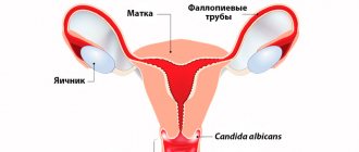
- Trichomonas vaginitis (trichomoniasis). Accounts for 15-20% of cases of vaginitis. Caused by the simplest single-celled microorganism Trichomonas vaginalis. It is transmitted sexually and affects the vagina, cervix and urethra, and also increases the risk of premature birth in pregnant women. With trichomonas vaginitis, the discharge has a greenish-yellow color and an unpleasant odor.
Who is at risk?
- Pregnant women.
- Women after menopause.
- Women who have multiple sexual partners.
- Women suffering from sexually transmitted infections.
- Women using oral contraceptives.
Diagnostics
If you have symptoms of vaginitis, you must consult a doctor who, after interviewing the patient, a gynecological examination and a number of additional laboratory and instrumental studies, will be able to determine the cause of the disease and prescribe effective treatment.
Laboratory diagnostics
- Microscopic examination of discharge from the genitourinary organs of women (microflora), 3 localizations. This is one of the main methods for detecting vaginitis. A sample of the material is examined under a microscope after preliminary staining with special dyes or without it. Allows you to identify signs of inflammation and detect the pathogen that caused the disease.
- Study of vaginal microbiocenosis with determination of sensitivity to antibiotics. The vaginal discharge is inoculated onto a specific nutrient medium, which makes it possible to detect even a small amount of the pathogen in the material. For vaginitis, a culture for mycoplasma, ureaplasma, chlamydia, Candida fungi, and trichomonas may be prescribed.
- Cytological examination of smears and scrapings from the surface of the cervix and external uterine pharynx - Papanicolaou staining (Pap test). This is a study of cells in the vagina and cervix to identify atypical, malignant cells. Necessary for diagnosing changes that precede cervical cancer. It is mandatory for women over 30 years of age and for women infected with certain types of human papillomavirus.
- Determination of specific immunoglobulins for pathogens of urogenital infections. It is carried out by enzyme-linked immunosorbent assay (ELISA). Allows you to identify antibodies (immunoglobulins) to certain infections - specific proteins that are produced in the body in response to the penetration of a microorganism. In this way, antibodies to yeast-like fungi, ureaplasma, and Trichomonas can be detected.
- Determination of the genetic material of the infectious agent in a woman’s vaginal discharge using the polymerase chain reaction (PCR) method. The DNA of chlamydia, mycoplasma, gardnerella and other pathogens is detected.
Other research methods
- Vaginal pH measurement. This is a study of the acid-base balance of the vagina. Normal vaginal pH is 3.8-4.2. To measure, use special indicator paper, which, upon contact with vaginal discharge, changes color depending on the pH level, or pH meters. With vaginitis, the pH may increase due to a decrease in the number of lactobacilli that form lactic acid.
- Aminotest. Used to diagnose bacterial vaginosis caused by Gardnerella vaginalis, it involves adding one or two drops of vaginal fluid to a 5-10 percent potassium hydroxide solution. The appearance of a fishy odor is a sign of gardnerellosis.
- Colposcopy. This is an examination of the vaginal walls using a special colposcope equipped with an optical system and a light source. Allows you to assess the condition of the vaginal mucosa, identify damage or neoplasms, and also perform a biopsy, that is, take a sample of tissue from the mucous membrane for subsequent microscopic examination.
If a patient is diagnosed with trichomonas vaginitis, the doctor may additionally prescribe tests for other sexually transmitted diseases. Sometimes additional studies are carried out to assess a woman’s hormonal levels.
Treatment
Treatment depends on the cause of vaginitis. Medicines can be used both in the form of suppositories, gels, ointments, and in the form of tablets or injections. The choice of drug, dose, and method of administration depend on the cause of vaginitis, the severity and duration of the disease, the presence of concomitant diseases and conditions. For example, antibiotics are used for bacterial vaginosis, and antifungal drugs are used for thrush. For atrophic vaginitis, a good effect is achieved by taking estrogen in the form of creams or tablets.
Prevention
- Compliance with the rules of intimate hygiene.
- Use of barrier methods of contraception.
- Timely preventive gynecological examinations.
Recommended tests
- Microscopic examination of discharge from the genitourinary organs of women (microflora), 3 localizations
- Culture of flora with determination of sensitivity to antibiotics
- Culture for Mycoplasma species with determination of titer and sensitivity to antibiotics
- Culture for Ureaplasma species with determination of titer and sensitivity to antibiotics
- Culture of Candida spp./yeast-like fungi with selection of antimycotic drugs
- Culture for Chlamydia trachomatis with determination of sensitivity to antibiotics
- Culture for Trichomonas vaginalis
- Study of vaginal microbiocenosis with determination of sensitivity to antibiotics
- Analysis of vaginal microbiocenosis. 8 indicators, quantitative DNA [real-time PCR] (urogenital smear)
- Analysis of vaginal microbiocenosis. 16 indicators, quantitative DNA [real-time PCR] (urogenital smear)
- Cytological examination of smears and scrapings from the surface of the cervix and external uterine pharynx - Papanicolaou staining (Pap test)
- Mycoplasma hominis, IgA
- Mycoplasma hominis, IgG, titer
- Chlamydia trachomatis, IgA, titer
- Chlamydia trachomatis, IgG, titer
- Chlamydia trachomatis, IgM, titer
- Chlamydia trachomatis, DNA [real-time PCR] (urogenital smear, urine, ejaculate, prostate secretion)
- Ureaplasma species, DNA quantitatively [real-time PCR] (urogenital smear, urine, ejaculate, prostate secretion)
- Trichomonas vaginalis, DNA [real-time PCR] (urogenital smear, urine)
- Trichomonas vaginalis, IgG, titer
- Candida albicans, IgG, titer
- Histological examination of biopsy diagnostic material (endoscopic material, tissues of the female reproductive system, skin, soft tissues)