When menstruation does not come according to the clock, it is difficult to calculate the exact date of the examination and appointment with the doctor. Is it possible to do an ultrasound during menstruation, will the results be correct - these questions are relevant for any woman. To be sure, it is important to understand the influence of the phase of the menstrual cycle on the state of the internal organs and systems of the body.
Proper preparation for the procedure
The influence of the regulator on ultrasound examination is as follows:
- The ability of ultrasound to penetrate tissue deteriorates. The most informative method for examining the reproductive organs is transvaginal ultrasound. It involves inserting a tube of the device into the vagina, in which blood accumulates, creating an additional barrier between the organs and the sensor.
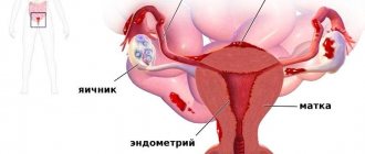
- The effectiveness of examining the uterine cavity is reduced. During menstruation, the uterus resembles an inflamed wound on which blood clots accumulate. The specialist will not be able to examine developing inflammation and small tumors.
- The actual thickness and structure of the endometrium cannot be assessed. During menstruation, the uterine layer actively peels off, but a new one has not yet grown.
In addition to the above factors, it is not advisable to perform an ultrasound during menstruation due to the fact that many women experience additional discomfort (abdominal pain, general malaise). Transvaginal examination also causes discomfort for the patient psychologically.
However, pelvic organs that are not related to the reproductive sphere can be examined during menstruation. Ultrasound of the bladder is done transabdominally, and examination of the rectum is done with a rectal probe.
During a routine examination of the reproductive organs, experts recommend doing an ultrasound examination on days 8–10 of the menstrual cycle. During this period, the discharge does not interfere with the examination of the uterus and vagina, and the endometrium is not yet dense and allows examination of medium-sized formations.
If it is necessary to carry out the procedure in the first days of the cycle, it also matters how many days have passed since the start of menstruation. The table shows recommendations regarding ultrasound depending on the day of the menstrual cycle.
| Type of procedure | Direction of research | Recommended day of the menstrual cycle |
| Transabdominal | Bladder | Any, if the woman is ready for the procedure |
| Transrectal | Rectum | |
| Transvaginal | Reproductive organs due to bleeding, pain, various complications | Day of admission to hospital |
| Myometrial examination | 1-3 | |
| Determining the type of tumor | 3-7 | |
| Functional ovarian cyst | 1-2 | |
| Monitoring follicle growth | 1-5 | |
| Diagnosis of endometrial hyperplasia | 1-3 |
Home » Kidneys » Is it possible to do an ultrasound of the kidneys during menstruation?
Ultrasound examination is the most common diagnostic method. In many situations, it is simply necessary to undergo an ultrasound urgently; this applies to those cases where there is a risk of having a disease or pathology that needs to be identified at an early stage. Women first of all try to rely on their menstrual cycle, believing that it affects the results of the study, let's try to find out if this is actually true.
If we are talking about ultrasound of the kidneys, heart, stomach, joints, thyroid gland and blood vessels, then there is only one answer - the female cycle will not affect the result in any way. And this procedure will not cause any harm, so there is no need to worry.
However, the situation is different with ultrasound of the pelvic organs. Blood clots located in the uterus at this moment simply will not allow the doctor to discern possible existing problems. The cyst will not be noticeable. A tumor or even a polyp if it has not yet reached a large size. In addition, this is an unpleasant procedure for both the doctor and the patient, because at this moment you will have to use a vaginal sensor, and for some patients this may even cause pain.
An ultrasound examination during menstruation will not harm a woman’s health, but in some cases it may distort the results. Therefore, do not be afraid of this safe procedure.
The main indications for a kidney ultrasound are detection of the following symptoms in the patient:
- diseases of the endocrine system;
- inflammatory processes in the body that are acute or chronic;
- pain in the lumbar region and lower abdomen;
- urinary disorders;
- formation of various benign and malignant formations;
- congenital anomalies of the external genital organs in women;
- changes in the functioning of the renal system;
- high blood pressure;
- swelling of the legs and puffiness of the face.
Proper preparation for an abdominal ultrasound: how not to spoil the study?
Ultrasound diagnosis of the abdominal organs has become a standard procedure in modern medicine. It allows you to conduct a full examination of the digestive system in a short time and detect inflammatory, degenerative, and oncological pathologies. But ultrasound requires special training, which can significantly improve its information content .
This article will answer the following questions:
- What are the dietary and drinking habits before an ultrasound?
- Is it necessary to do an enema or take special medications ?
- Are there any special preparations for a child or woman during menstruation ?
- What do you recommend taking with you to the study?
- What is the price of abdominal ultrasound in public and private clinics?
What internal organs are examined?
During an abdominal ultrasound, the following organs are examined:
- liver;
- gallbladder and bile ducts;
- spleen;
- stomach;
- small or large intestine;
- pancreas.
In addition to the digestive system, during the study, large vessels (abdominal aorta, portal vein of the liver), and lymph nodes are examined. Sometimes the diagnosis is supplemented by examination of the kidneys, adrenal glands and organs of the female reproductive system (uterus and its appendages).
Principle of nutrition before the procedure in adults
The presence of food masses in the stomach and intestines significantly reduces the effectiveness of diagnostics. Therefore, all adult patients are advised to refrain from eating 6-8 hours before the ultrasound .
It is also necessary to take into account that increased gas formation in the intestines negatively affects information content. Therefore, patients with this problem (mostly elderly people with chronic pathologies) are prescribed a diet for several days. All products that may contribute to the formation of gases are removed.
Help You can learn more about the diet before an abdominal ultrasound from a separate article, where we looked at what you can and cannot eat before the procedure and gave an example of a menu for three days.
No attention is paid to nutrition before an ultrasound if the patient needs an examination as soon as possible (for example, with acute abdominal pain).
If you delay time in such a situation, it can negatively affect the patient’s health.
Taking medications
Before the study, the following drugs are prescribed:
- Sorbents (activated carbon, Smecta) are a group of medications that are capable of absorbing and removing toxic substances and gases from the lumen of the digestive system. Prescribed if the patient has a tendency to flatulence, on the day of the ultrasound. It must be taken into account that they contribute to the development of constipation.
- Espumisan (simethicone) is a pharmacological agent that is used for increased gas formation in the intestines. After use, simethicone destroys air bubbles, which facilitates its removal from the digestive tract. Appointed on the day of the study.
- Laxatives. In some patients with chronic constipation, stool fills the lumen of almost all parts of the large intestine. This reduces the transmission of ultrasonic waves, which worsens the quality of diagnosis. Therefore, they are preliminarily (24 hours before) prescribed laxatives (macrogol), which help to completely cleanse the digestive tract.
- Analgesics and antispasmodics. Patients with gastrointestinal problems often experience pain, sometimes of a spastic nature (for example, with cholelithiasis or intestinal colic). To reduce it, adequate pain relief is necessary before the procedure. For this purpose, non-steroidal anti-inflammatory drugs (ibuprofen, paracetamol) and/or antispasmodics (drotaverine) are given 30-40 minutes before the start.
- Other medications. Sometimes additional medicinal preparation of the patient is required. For epilepsy, anticonvulsants are prescribed, and for severe fear, sedatives are prescribed.
Quite often, patients ask the doctor whether it is necessary to do a cleansing enema before an ultrasound of the abdominal organs. If you plan to carry out a standard diagnostic procedure ( through the anterior wall of the abdomen ), then there is little point in doing it.
The fact is that during an enema, feces are removed only from the rectum and sigmoid colon , which does not particularly affect the information content of the examination of other areas of the digestive system. It is rational to do it if a transrectal (through the rectum) ultrasound or endoscopic examinations (colonoscopy, irigoscopy) are planned.
How should a woman prepare for an examination?
Menstruation or the monthly cycle also do not affect the information content of the examination of the digestive system. However, they are important if additional routine diagnostics of the uterus and its appendages (pelvic ultrasound) is planned. It is recommended to carry out it on days 5-7 of the cycle (in the stage of early proliferation), when changes in the walls of organs are most easily detected.
An ultrasound also does not require a woman to stop breastfeeding. Ultrasonic waves do not affect the activity of lactation, the quality and quantity of milk.
Often pregnant patients are afraid to undergo abdominal ultrasound because they believe that it can harm the development of their child. But numerous studies have shown this statement to be incorrect. On the contrary, ultrasound imaging methods are called the safest for the expectant mother, since they do not carry an ionizing or magnetic resonance load.
Reference: In 2009, an analysis was carried out of 55 studies from around the world that monitored children during pregnancy and after birth until they were 8-9 years of age.
The authors (Laura Houston, Antony Odido, George Macones) concluded that ultrasound during pregnancy does not affect the development of the child or the presence of health problems.
On the contrary, it promotes early diagnosis of diseases, which reduces mortality and miscarriage rates (https://obgyn.onlinelibrary.wiley.com/doi/abs/10.1002/pd.2392)
Is it possible to do research for children?
Preparing children for ultrasound diagnostics has several important nuances that you should always pay attention to:
- Psychology. It is necessary to talk with the child first and convince him that the procedure is absolutely safe and painless. At the same time, it is important for parents to maintain external calm.
- Shorter duration of abstinence from food (depending on age - 2-6 hours).
- Creating a comfortable environment. Parents are advised to bring toys with them to create a home environment for the child.
- Maximum courtesy and understanding from the medical staff.
Memo for the patient
The following table contains answers to the most common questions from patients regarding whether it is possible to perform a certain action before performing an ultrasound of the abdominal organs.
| Is it possible before performing ultrasound diagnostics? | |
| Smoking | Not recommended. May affect the functioning of the digestive system |
| Tea | You can drink weak tea 1 hour before starting the diagnosis. |
| Coffee | It is not advisable to drink coffee 24 hours before an ultrasound. |
| Kefir | Equates to food. Do not drink for 6-8 hours before the procedure |
| Alcohol | Affects the functioning of the digestive system (pancreas, liver, stomach). Do not consume within 3 days before ultrasound |
| Teeth cleaning | Allowed |
| Medicines | All medications prescribed for the treatment of chronic pathologies are taken as usual |
| Bananas | It is impossible 6-8 hours before the start of the ultrasound. |
| Drinking water | Allowed (even immediately before the study) |
What to take with you?
Doctors always advise taking food with you for the study. Since the patient is forced to fast for up to 8 hours, immediately after completing the diagnosis, you can snack on croissants, sandwiches or fruit.
It is advisable to come to public clinics (especially in small towns) with your own towel, which can be used to remove any remaining gel.
It is important to remember to take medical documentation. If the patient is inpatient treatment, this is the medical history; if not, then the patient’s outpatient card. It (if available) must be accompanied by a referral from the attending physician, an insurance policy and documents that confirm the patient’s identity (passport or birth certificate).
Please note: The results of previous examinations (ultrasound, CT, MRI) of the abdominal organs will be informative for the doctor who will conduct the diagnosis. This will allow him to compare the results obtained with previous ones.
Price
In Moscow, abdominal ultrasound is performed in numerous hospitals, clinics and diagnostic centers. The cost of the procedure in private medical institutions is 1200 (MedFord) - 6200 (DR Medical Group) rubles, and in public hospitals - 1500-3200 rubles.
It is not difficult to get examined in St. Petersburg . Its price in private centers is from 1,200 rubles (“AlfaMed”, “Blagodatnaya”, “Dynasty”). Immediately after the ultrasound, you have the opportunity to consult with a specialized specialist.
Diagnosis of abdominal organs is somewhat cheaper in the regions . In Yekaterinburg its cost is 900-1200 (“Medline”, “Angioline”), in Kazan – 550-1500 (“MEDEL”, “Remedy”, “Nashe Delo”), in Nizhny Novgorod 750-1500 rubles (“Svet”, "Health", "Euroclinic").
You can undergo a free abdominal ultrasound if you have an insurance policy (which covers this diagnostic method) and a referral from your attending physician (not older than 3 months). This information must be provided when registering the patient for the study.
Conclusion
Proper preparation for an abdominal ultrasound can ensure a highly informative diagnostic process. Its components are proper diet, medication, and psychological work. Special preparation is not required only if the patient enters the clinic with an acute pathology or emergency condition.
Source: https://gastrosvoboda.com/diagnostika/uzi/podgotovka-k-protsedure.html
Kidney function and menstruation: why kidneys hurt during menstruation, reasons for delay, kidney ultrasound
A woman's genitals do not function autonomously. Their work is also influenced by other systems. The genitourinary organs located near the uterus and appendages are capable of influencing them. The functioning of the kidneys and the course of critical days are interdependent processes. The article talks about this in detail.
Removing waste products is not the only function of the paired organ. It is also involved in the functioning of the endocrine system, namely in the production of the hormones erythropoietin, renin, and prostaglandin. The first is responsible for the production of red blood cells, the second for fluid exchange and blood circulation, the third for the normal size of the lumen of blood vessels and blood pressure. This confirms the influence of the kidneys on menstruation.
Menstruation is possible only under the influence of certain factors, in the implementation of which the paired organ plays an important role. Among them the following are noted:
- stabilization of acid and alkaline components in the body;
- metabolism, that is, the transition of one substance to another;
- development of the reproductive system, normalization of hormonal levels;
- improvement of the condition of blood vessels, synthesis of blood components, part of which is consumed during menstruation.
Any deviation in the functioning of the urinary system has a negative impact on health.
Is it possible to do an ultrasound of the kidneys during menstruation?
Ultrasound examination of a paired organ is a painless, safe and performed in a short period of time method for diagnosing a number of pathologies of the urinary organs. With the help of ultrasound, the doctor will be able to identify their size, structure, location, assess the condition of the vessels, and detect abnormalities, for example, stones or neoplasms.
Critical days are not included in the list of contraindications for ultrasound. The study does not affect the hormonal system and women's health in general.
Indications for ultrasound examination during menstruation
Despite the complexity of the procedure, there are situations when research is still carried out during menstruation:
- It is necessary to examine the middle uterine layer. The muscular layer of the uterus (myometrium) is clearly visible when the endometrium is thin, which is what happens during menstruation.
- Heavy bleeding occurs during menstruation. Since intense bleeding poses a threat to a woman’s life, ultrasound is performed regardless of the day of the menstrual cycle.
- The type of tumor is determined. Malignant tissues are clearly visible on the first day of the monthly cycle.
- My period started after pregnancy was confirmed. In this case, spotting serves as a signal about the pathology of pregnancy. The procedure will allow you to determine the tactics of further therapy.
- A woman has been diagnosed with a functional ovarian cyst. Ultrasounds are done on different days of the cycle to track changes in the size of the formation.
- The cause of infertility is being determined. For diagnosis, it is necessary to observe the maturation of the follicle.
- A woman is worried about intense pain in the abdominal area, the causes of which are unknown.
- Complications arose after insertion of a device into the uterus, surgery on the reproductive organs, abortion or miscarriage.
- Menstruation is accompanied by nonspecific symptoms (purulent discharge, unpleasant odor).
Timely examinations are necessary, because very often all kinds of disorders of internal organs sometimes pass without obvious manifestations: sometimes there is no pain or even discomfort. This is due to the fact that there are fewer nerve endings in the abdominal cavity and pelvic organs than in other parts of the body (a natural mechanism that protects a woman from unbearable pain during childbirth). Therefore, prevention is important, and it begins with an ultrasound examination.
Ultrasound is one of the best and most effective ways to study internal human organs. Ultrasound examination is very easy to perform, harmless, painless, effective and absolutely time-consuming.
For the health of any woman, one of the most significant examinations is an ultrasound examination of the pelvic organs.
Ultrasound examinations can be performed either spontaneously or planned. Most often, women make an appointment with a doctor several weeks in advance. Of course, it’s good if everything goes well and the doctor’s appointment is planned and does not coincide with the menstrual cycle. But there are situations when one overlaps with the other. And here most representatives of the fair half have completely natural questions:
- Is it better to do an ultrasound during menstrual flow or is it safer to delay this procedure?
- What is the procedure for dealing with the current situation?
- At what point should an ultrasound of the female genital organs be done during menstruation?
- In what situations do doctors not recommend such a procedure?
Ultrasound during menstruation: is it acceptable?
An ultrasound of the abdominal cavity during menstruation is definitely possible. Do not worry that vaginal discharge will interfere with the doctor’s performance of the procedure. There are a number of procedures where this fact will indeed be an obstacle. Ultrasound is not included in this number. Doctors are aware of what to do in such situations, and therefore they themselves will navigate the optimal prescriptions. If it is necessary to diagnose the pelvic organs, the study is prescribed in the first part of the monthly cycle, but not earlier than the first day.
Breast ultrasound
Breast sonography does not use X-rays or other potentially harmful types of radiation. It is often used to image breast tissue in women of reproductive age. It is especially valuable when it is necessary to distinguish a cyst (a fluid-filled sac) from a solid formation (filled with tumor masses).
Which days of the cycle are most suitable for breast examination? It is better to do an ultrasound after menstruation, from 7 to 14 days of the cycle. The mammary glands are a hormone-dependent organ, therefore, during reproductive age, cyclical changes in tissues occur in them, periods of increased density are replaced by decreased density, which affects the information content of ultrasound examination.
Indications for use
Ultrasound scanning of the breast typically complements mammography and magnetic resonance imaging (MRI). Ultrasound of the mammary glands is prescribed to:
- find the cause of pain, swelling and redness in the chest;
- evaluate tumor-like formations detected as a result of self-examination or clinical examination of the breast;
- obtain additional information when identifying abnormal changes in breast tissue detected during mammography;
- control the advancement of the needle when performing a breast biopsy, drainage of a cyst or abscess of this organ;
- monitor changes in the size of cysts or benign tumors (fibroadenomas);
- determine how far the breast cancer has spread;
- examine the mammary glands if a silicone implant is installed, since mammography in this situation is not very informative.
We recommend reading the article about breast ultrasound. From it you will learn about the possibility of carrying out the procedure on critical days, the recommended period for examination, and the methodology for carrying out this procedure.
How to prepare for an ultrasound?
Regardless of the field and method of research, the following principles must be adhered to:
- 2-3 days before the procedure, exclude foods that cause increased gas formation from the diet;
- on the eve of the study, have a bowel movement or an enema;
- Ultrasound should be performed in the morning.
Women prone to flatulence are advised to take medications that eliminate the problem (for example, Espumisan) for several days. When performing a bladder examination, as well as a transabdominal examination, the bladder must be filled. To do this, 2-3 hours before the ultrasound you need to drink 1.5–2 liters of water and refrain from urinating.
During a transvaginal examination, the bladder must be empty. Before examination with a vaginal sensor, you also need to take care of a clean diaper and a spare pad.
On what day of the cycle should I do a gynecological ultrasound?
Gynecologists have a common recommendation for all women: visit a doctor and sign up for an ultrasound examination from days 5-7 to 10-11 of the cycle. Each individual case requires individual recommendations.
On what day of the cycle should I do an ultrasound of the uterus?
If there is a suspicion of the development of fibroids, an ultrasound is performed on the 3rd day of menstruation or immediately after its end. If fibroids, fibroids, or polyps are identified earlier, ultrasound scanning is performed during menstruation. To accurately diagnose endometriosis, ultrasound examinations are performed before and after menstruation. Thus, it is possible to more accurately estimate the thickness of the inner layer of the uterus.
On what day of the cycle should I do an ovarian ultrasound?
The optimal time for such a study is days 5-7 of the cycle. This applies to the diagnosis of ovarian pathologies and monitoring when they are identified earlier. If folliculometry is necessary, then ultrasound monitoring is carried out starting from the 10th day of the cycle every 2-3 days.
On what day of the cycle should I do an OMT ultrasound?
It is optimal to perform an OMT ultrasound on days 5-7 of the cycle. The study of the pelvic organs refers to the assessment of the structure and condition of:
- uterus,
- appendages,
- Bladder,
- cervix,
- vagina.
For this period, it is possible to exclude the formation of functional cysts, which disappear by the end of menstruation. At the moment, it is easier to evaluate polypous formations. In special cases, a pelvic ultrasound can be done during menstruation, without waiting for it to end.
Features of performing an ultrasound of the pelvic organs during menstruation
The main feature of ultrasound of the pelvic organs during menstruation is that it is performed only if there are specific indications or in emergency situations. If the examination can be postponed, the doctor will schedule the procedure for another day.
Despite the unhygienic nature of manipulations during menstruation, transvaginal examination is the preferred method for ultrasound of the pelvic organs. However, during menstruation, the cervical canal opens slightly, and the danger of pathogenic microorganisms entering the uterine cavity increases. In this regard, transvaginal ultrasound is not performed during the period of regulation if a woman has infections in the vagina.
Another feature of ultrasound during menstruation is that during the examination, many women also experience pain in the lower abdomen. Discomfort occurs due to the fact that the uterus becomes very sensitive during this period. Moving the device's sensor causes spasms, which contribute to the appearance of unpleasant sensations.
Cystitis during menstruation
This disease is the result of inflammation of the mucous membrane of the bladder and is more common in women. Many representatives of the fairer sex know that cystitis also occurs during menstruation. Why is this happening? What does it indicate? How is the disease treated on days like these?
Features of the disease during menstruation
Answering the question about cystitis during menstruation, doctors explain that this is due to inflammatory processes in the pelvis. And this is no longer just an annoying misunderstanding, but a rather serious problem. Therefore, you should not use improvised means for pain relief or relief from the symptoms of the disease. It is better to see a doctor to prescribe the correct treatment. If this is not done, the disease will become chronic and may contribute to the development of pyelonephritis.
Inflammation of the genitourinary system also provokes a decrease in bladder volume. And this can no longer be done without surgical intervention.
An infection in the bladder does not just appear during menstruation. The process begins in the uterine appendages under the influence of gram-positive bacteria that enter there. Further development of the infection takes on a latent form.
When menstrual bleeding begins, the body naturally cleanses itself. Bacteria leave their habitats. They move into the urethra, entering the urethra into the bladder. This phenomenon is often accompanied by severe pain. Such a manifestation of the disease is a signal of algodismenorrhea, that is, dysfunction of the genital organs. Its symptoms also include: nervous tension, pain when urinating, increased frequency, irritability, heaviness in the lower abdomen.
Exceptions in which examination is prohibited
Sometimes menstruation is not a reason to refuse an ultrasound. In medical practice, there are situations when, despite bleeding, information about the condition of certain organs is urgently needed. The doctor may urgently order an ultrasound examination for the health and safety of the patient. And then it is still necessary to perform a pelvic ultrasound even during menstruation. Such situations include:
- heavy bleeding;
- severe, prolonged pain of unknown origin;
- suspicion of ectopic pregnancy or threat of spontaneous abortion;
- suspected pathology (uterine fibroids, hyperplasia, ovarian cyst).
When screening an ovarian follicle, it is important to perform several stages of ultrasound over 1–15 days of the menstrual cycle. Patients who experience increased flatulence should exclude all foods that cause intense gas formation from their diet 2-3 days before the procedure. These include all sweet foods, laxatives (legumes, cabbage), fruits, flour products. In addition, it will be useful to take an enema before the upcoming examination.
The presence of blood in the uterus does not prevent either the visualization of the fertilized egg or the determination of the strength of its attachment to the wall. There are also cases with various diseases of the female organs, when it is especially important for doctors to conduct an examination precisely at the initial stage of the menstrual cycle.
Possible causes and treatment of thickening of the bladder walls
IT IS IMPORTANT TO KNOW! The only remedy for CYSTITIS and its prevention, recommended by our subscribers! Read more…
Very often, when examining women, doctors are faced with such a pathology as thickening of the bladder wall. The disease can be accurately determined using ultrasound. The data obtained after diagnosis are analyzed and, based on the results, a final diagnosis is made, the causes of thickening of the bladder walls are determined and treatment is prescribed.
What symptoms warrant an ultrasound?
Knowing what signs indicate the onset of the disease, the patient has every chance to stop the development of the disease at an early stage.
- Frequent urination in women, accompanied by pain and discomfort in the lower abdomen. This is not the norm. Such symptoms can become signs of an inflammatory process in the bladder, urolithiasis, accompanied by the passage of stones, and malignant formation of the bladder.
- Hematuria – there is blood in the urine. The process of urination is accompanied by a burning sensation. These signs may be a signal of urolithiasis, the development of a tumor or injury in the area of the genitourinary system.
- Feeling that after emptying the bladder remains full.
- When examining urine, the following abnormalities are found: protein and microorganisms, increased levels of casts and leukocytes.
Very often, during cystoscopy (the main method of examining the bladder cavity), a polyp, malignant formation and other pathologies can be identified.
If thickening of the bladder walls is diagnosed, it is necessary to find the reasons that led to this disease.
Thickening: general and local nature
General reasons that led to general thickening.
- In the male half, these may be obstructions located in the ducts of the prostate gland (the ducts are clogged with stones coming out of the kidneys or bladder). Such neoplasms require the patient to undergo an ultrasound of the prostate gland as soon as possible.
- Hydronephrosis is an enlargement of the renal pelvis. The pathology is of an increasing nature; one can be born with it or acquire it during one’s life. In this case, the patient is prescribed diagnostics of the ureters and kidneys.
- Bladder diverticula are protrusions in the form of pouches. With this pathology in women and men, urination is carried out in two stages: first the bladder is emptied, and after that the urine comes out of the bags. When the second part of the process does not occur, disease may develop.
- Chronic inflammation of the bladder. In this case, a dense, uneven formation with blurred contours appears on the wall of the bladder.
- Schistosomiasis is an infectious pathology caused by the presence of microorganisms. Thickening in this case in women and men occurs due to calcium deposition.
Local causes of wall thickening.
- Wrinkling caused by insufficient filling.
- Malignant cancers of the bladder. They don't cause pain.
- Tuberculosis, in which granulomas form (inflammation of tissue that closely resembles small nodules). They are what lead to the appearance of local thickenings.
- Pelvic injuries, and their consequence is the formation of a hematoma on the bladder.
- Polyps. They can be either very small or very large.
To understand whether there is a pathology, you need to know exactly what the normal thickness of the bladder wall is.
Dimensions of the bladder walls: normal
After the diagnosis, only the doctor should decipher it so as not to miss malignant neoplasms and other pathologies. If no diseases are detected, then the characteristics of the bladder should be as follows.
- The shape of the organ is smooth and round, the norm on longitudinal scanning is ovoid.
- The contours inside and outside the bladder are clear and even.
- The normal wall thickness is within 3 - 5 mm (in an adult); it should also be remembered that the more filled the organ is, the thinner its walls.
- Urine flow is 14 cm per second, this is normal.
- The remaining urine should not exceed 50 ml.
If, nevertheless, the norm is violated and the patient has a fine pathology, suspension in the bladder, inflammation, malignant neoplasms, schistosomiasis and other pathologies, then the doctor prescribes treatment. It will depend on the diagnosis.
Treatment with drugs
First of all, before removing the suspension in the bladder, you need to relieve inflammation. During an exacerbation or if there is severe pathology, malignancy or other diseases, it is better for the patient to stay in a hospital under the supervision of specialists. Drug treatment is aimed at eliminating the infection and inflammatory process, therefore broad-spectrum antibiotics are prescribed, the duration of which is from 14 days, depending on the complexity of the disease.
Also, thickening of the walls involves taking other drugs, such as Allohol, Cholenzym, Nicodem, Oxafenamide. If the pathology is sluggish, then cholekinetics are prescribed. But these drugs should be taken with caution by those patients who have gallstones. The following drugs showed very good results in therapy: Atropine, Papaverine, Eufillin, Nitrite, Amizil.
If a tumor is detected in a patient, then surgery and chemotherapy cannot be avoided. But in any case, treatment is selected individually for each patient, depending on his disease and concomitant pathologies.
Treatment using traditional medicine methods
Not only drugs are effective in the treatment of wall thickening, but also traditional medicine gives good results, even if the formations are malignant. The following recipes are effective.
- Mix 2 parts of mint leaves, 4 parts of dandelion roots, 1 part of celandine, 2 parts of toadflax and the same number of tansy flowers, 4 parts of cinquefoil roots. 1 tbsp. collection, pour 250 ml of water, infuse for half an hour and take three times a day before meals.
- Take 500 ml of beet juice, carrots, aloe, black radish, vodka and honey. Everything is poured into one jar, mixed well, rolled up with a lid and buried in the ground for two weeks. After the jar is dug out, the resulting product is taken ½ cup three times a day.
There are many recipes, each of them is good in its own way, and some can completely stop the growth of the tumor. Before using this or that recipe, it is better to consult a doctor so as not to harm your health, because there are also methods that can provoke intensive growth of the tumor.
By secret
- Incredible... Chronic cystitis can be cured forever!
- This time.
- Without taking antibiotics!
- That's two.
- During the week!
- That's three.
Follow the link and find out how our subscribers do it!
×
Diagnosing the ovaries
For menstrual flow, there are 2 ways to perform ultrasound examinations. They differ in the principle of implementation. These include:
- transabdominal method;
- transvaginal.
Often, in order to get a complete picture of the condition of a person’s genital organs, both methods are used.
The transabdominal method is external, less labor-intensive and time-consuming. The doctor moves the meter along the visible part of the patient’s abdomen, having previously smeared it with medicinal gel. All indicators of the condition of the ovaries are displayed on the monitor.
The most accurate indicators are produced by the second method - transvaginal. When using it, the sensor is placed inside the girl’s body through the vagina.
To maintain the required level of hygiene and for the comfort of the examinee, a gel-coated condom is placed on the sensor. Such an ultrasound does not last long and produces more accurate results. With good professional training of the medical worker, the patient does not experience severe discomfort.