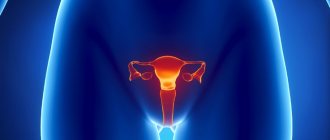
A woman's health is very fragile. Doctors often diagnose cervical pathologies in women that are associated with the inflammatory process or are their consequences - erosion, dysplasia, ectopia, as well as cancer. Modern medicine offers a unique method of therapy called conization. The essence of this procedure is to remove a cone-shaped area of the surface of the cervical canal or a portion of damaged muscle tissue.
Conization of the cervix is one of the methods for treating precancerous diseases and preventing cancer. The operation is low-traumatic and does not require hospital treatment. Often the procedure is performed not for treatment, but for the purpose of research and diagnosis of the underlying disease. A woman’s well-being after cervical conization is determined by many factors. The recovery period proceeds strictly individually for each patient and directly depends on concomitant diseases, the state of immunity, as well as on the chosen method of medical intervention.
Postoperative period
After conization of the cervix, patients do not experience any pain other than mild discomfort. Rehabilitation is outpatient. If the operation was performed with a laser or using the radio wave method, then the likelihood of complications is minimal. In the presence of acute pain, as well as against the background of severe uterine bleeding or high fever, you should immediately seek medical help.
It takes about a month for the organ to heal completely. What contraindications should you be aware of after cervical conization? It is required to take a break from sexual activity, canceling trips to the bathhouse, swimming pools and sauna at the same time. Another rule of rehabilitation is the limitation of any physical activity.
Types of surgery
Modern medicine has several methods of surgical treatment. The method of conization of the reproductive organ is selected individually for each woman. The essence of the available surgical methods is presented below.
Surgical
This type of surgical treatment is currently used for special indications. Surgical excision of diseased tissue on the cervix has many disadvantages, such as a high rate of trauma, the risk of infection, and severe blood loss. The advantage of this treatment method is the high accuracy of removal of affected tissue.
Laser
This method is quite popular, but also not widely used. In this case, the high cost of the operation played an important role, as well as the need for general anesthesia for the patient. Such a surgical intervention involves excision of diseased tissue with a laser beam or evaporation. The disadvantages of the presented treatment method are high cost and general anesthesia. The advantages include high precision of excision and high-quality therapy.
Electrowave
Electrowave conization - otherwise called radio wave - is the most popular method of cervical conization in clinics. Removal of affected tissue is carried out using an electrode loop to which alternating current is supplied. Electroconization has many advantages:
- low cost;
- no unpleasant consequences;
- local anesthesia;
- little blood loss.
The disadvantages of treatment include the inability to control the depth of excision.
Cryoconization or freezing
The use of nitric oxide leads to freezing of the diseased tissue area, after which the lesion is destroyed. This method is painless and not too expensive, but is currently almost never used in the country. This factor is explained by the inability to calculate the power of the freezing factor, as well as the subsequent lack of a piece of tissue for analysis. This treatment method cannot be used if cancer is suspected.
After colonization
What does the cervix look like after conization? Restoration of the organ is characterized by the following points:
- A deep wound is formed in the area of the removed tissue, accompanied by bleeding in the first day after conization.
- Slowly, as it heals, the wound is usually covered with a scab if the operation was performed using radio waves or laser methods.
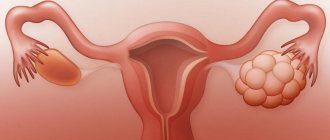
- Directly below the scab, active healing is observed. Then, at a certain point in time, it can separate from the cervix and come out naturally. This process is often accompanied by resumption of bleeding. As a rule, separation occurs after ten to fourteen days. But this period is individual and is determined by the size of the removed lobe of the cervix after conization. The photo shows a diagram of such manipulation.
Features of rehabilitation
Any surgical intervention in the anatomy of this organ entails a long recovery period. Basically, recovery after conization of the cervix depends on the behavior of the patient: taking medications prescribed by the doctor along with hygiene, refusing sexual intercourse, adhering to an exercise regimen, and so on. The stages of therapy include conservative treatment aimed at accelerating the healing process, as well as preventing relapses of the disease.
The operation is performed only with the help of sterile instruments, but the risk of infection after it still remains. To minimize this likelihood (infection), the doctor may prescribe a course of therapy with antiseptic suppositories. It is in order to prevent infectious penetration into the wound that women must maintain absolute sexual rest, avoiding bathing and visiting swimming pools. The postoperative period does not require treatment of the damaged surface. It is unacceptable to douche yourself. It is necessary to ensure absolute physical rest of the damaged area.
Radio wave and laser techniques, as well as diathermoconization, allow the formation of a scab. Its release may be accompanied by increased bleeding. Once the surgical site is completely cleansed, the discharge will soon decrease.
The patient should be concerned about factors such as an increase in the abundance of discharge of an uncharacteristic type (presence of a cheesy consistency and yellow color), a sharp unpleasant odor. Such signs may indicate the emergence of bacterial flora. Doctors consider a slight feeling of pain, which is similar to premenstrual syndrome, to be acceptable. Doctors in the postoperative period, as a rule, prescribe women to take mild painkillers. So, below we will tell you what medications you can take after conization of the cervix.
What medications are used during the recovery period?
Since conization surgery involves removing the area of affected tissue, the regenerative process may take longer than initially expected. To speed up healing, drugs like Panthenol, Methyluracil, Levomekol and other drugs are used.
If the patient experiences itching or burning, this may be a symptom of an infection. Discomfort can be accompanied by an increase in temperature, and, at the same time, increased discharge. Preventive therapy in the postoperative period may include the following antimicrobial suppositories, for example, Hexicon along with Terzhinan and Rumizol.
Negative aspects in the postoperative period
Any surgical intervention and therapy for cervical diseases can lead to serious consequences. Complications, as a rule, arise in the conditions of a poorly performed operation, due to the complexity of the woman’s initial disease, as well as due to her non-compliance with the proposed recommendations. The main negative aspects of conization during the postoperative period are represented by the following factors:
- The occurrence of bleeding (about five percent of operations have these consequences).
- Development of an infectious and inflammatory process.
- The appearance of pain syndrome.
- The presence of scarring and stenosis.
- The occurrence of isthmic-cervical insufficiency (ICI) during pregnancy, causing spontaneous miscarriages or premature birth. It is worth noting that ICI does not always develop in patients after conization. Considering that its cause is a hormonal imbalance, along with a congenital disorder in the ratio of the connective tissue and muscle components, the operation may not affect the pregnancy process.
It is not always possible to feel good after conization of the cervix. Sometimes complications arise.
Pregnancy and childbirth after radioconization
Unlike other treatment methods, after radioconization, stenosis of the cervical canal does not occur and complications that can affect the course of pregnancy and childbirth do not develop. The cervix heals without scarring. The cervical canal remains patent and sperm can fertilize the egg without interference. After the therapy, a woman can conceive a child. It is recommended to plan pregnancy 2-3 months after surgery.
It is important to know
Properly performed radioconization does not interfere with conception, pregnancy, or natural childbirth.
Radioconization of the cervix does not affect the course of pregnancy and does not interfere with the development of the fetus. Since scars do not form on the cervix, the woman is not at risk of developing isthmic-cervical insufficiency. In the absence of other complications, a successful pregnancy and birth of a child is possible.
Childbirth after surgical treatment occurs without complications. A woman can safely give birth to a child through the vaginal birth canal. Radio wave treatment is a gentle method that does not have a negative effect on the cervix, does not interfere with its stretching during childbirth and does not interfere with the birth of the baby.
The successful course of pregnancy and childbirth largely depends on how the postoperative period went. If a woman followed all the doctor’s recommendations and was regularly observed by a gynecologist, she has a very high chance of experiencing all the joy of motherhood without significant problems and complications.
Interesting video about the benefits of radio wave conization
What to do if the uterus is bleeding?
Many women complain that the uterus bleeds after conization of the cervix. Such a complication during the postoperative period is usually associated with damage to the vascular system of the organ. Against the background of a violation of the clotting process, blood clots form. In addition, due to the discharge of a large scab, heavy bleeding may occur, which will require a visit to a doctor. The occurrence of insignificant discharge during the recovery process is a completely acceptable manifestation. This is due to a failure in the integrity of the walls of the organ. The discharge is bloody at the initial stage, and then becomes bloody.
Dysplasia after conization of the cervix
Often the reason for a doctor to prescribe conization of the cervix is the discovery of dysplasia. The goal here is to study the obtained biological material for the presence of malignant processes and remove the pathology as such. In some situations, removal of an area of the mucosa that has been affected by dysplastic processes is quite sufficient for treatment.
Cervical dysplasia in patients is characterized by atypical cells on the organ. The goal of treating this disease involves minimizing the risk of the disease progressing to the cancer stage. The main cause of dysplasia in women is considered to be infection with the papilloma virus. Therapy largely depends on the extent of the disease, the patient's age and her reproductive plans. Without treatment, dysplasia is likely to transform into cancer. In some cases, after conization, a recurrence of dysplasia is detected.
Pregnancy in women after treatment of cervical intraepithelial neoplasia
One of the pressing problems in obstetrics and gynecology is the problem of reproductive function in women after the use of invasive methods for the treatment of cervical intraepithelial neoplasia (CIN). In recent years, there has been a negative trend in the increase in the incidence of cervical cancer in young women. Therefore, pressing issues include not only early diagnosis of cervical CIN, but also timely organ-preserving treatment.
Table 1. Complications of the first and second trimester of pregnancy in patients after invasive methods of treatment for CIN of the cervix
Drawing. Methods of treatment of cervical intraepithelial neoplasia in the history of the examined patients (%)
Table 2. Complications of pregnancy in the examined patients in the third trimester (n = 50)
Table 3. Pregnancy outcomes in women after treatment for CIN
Adequate treatment of preinvasive carcinoma (severe dysplasia) of the cervix in women of reproductive age, according to modern concepts, is conization of the cervix, which implies both a diagnostic and therapeutic procedure (4).
No less important is the issue of the occurrence of relapses during pregnancy and after childbirth in women who have undergone invasive treatments for cervical precancer (1, 5).
The pregnancy rate after invasive methods of treating cervical diseases varies, according to various authors, from 15.9 to 36.7% (2, 5). There are conflicting opinions in the literature about the course of pregnancy and childbirth after destructive and invasive treatment methods.
Thus, according to M. Kasum (1991), the frequency of urgent births in patients after cone-shaped excision of the cervix is 66.2%, premature births - 14.7%, spontaneous abortions - 19.1% (8).
According to Bulgakova S.V. (2007), the frequency of spontaneous abortion in patients after cervical amputation was 20.3%, artificial termination - 34.4%, surgical birth - 14.1% and vaginal birth - 29.6% (2).
In this regard, the purpose of this study was to determine the characteristics of the course of pregnancy and childbirth in patients after invasive methods of treatment of cervical intraepithelial neoplasia of the cervix.
Materials and methods
To achieve this goal, we analyzed the course of pregnancy and childbirth in 50 pregnant women after invasive methods of treating CIN.
22 (44%) pregnant women had a history of cervical intraepithelial neoplasia II (CIN II), which corresponds to a moderate degree of dysplasia, of which 10 (20.0%) patients underwent diathermocoagulation (DEC) and 12 (24.0%) – radiocoagulation.
28 (56%) patients were diagnosed with cervical intraepithelial neoplasia III (CIN III), including severe dysplasia 20 (71.4%) and carcinoma in
situ
8 (28.6%). Of these, 21 (42.0%) patients underwent conization of the cervix and 7 (14.0%) underwent amputation of the cervix (see figure).
All pregnant women underwent a comprehensive examination, including general clinical, microscopic, bacteriological examinations, detection of urogenital infections, including human papillomavirus (HPV) and its highly oncogenic strains, using polymerase chain reaction (PCR), extended colposcopy, cytological examination of smears from the exo- and endocervix, ultrasonography.
Results and discussion
The average age of the examined patients with a history of CIN II was 26.5 ± 3.2 years and with CIN III – 30.2 ± 4.5 years. When analyzing reproductive function, it was revealed that this pregnancy was the first in 4 (18.2%) pregnant women with CIN II and only in 3 (10.7%) patients with CIN III. 18 (36.0%) women had a history of miscarriages.
In 2 patients with CIN III, pregnancy occurred after the use of assisted reproductive technologies. The first birth was expected in 17 (34.0%) pregnant women.
The period of time that elapsed after treatment for cervical intraepithelial neoplasia until the onset of actual pregnancy ranged from 2 to 6 years.
Considering the etiological role of PVI in the development of precancer and cervical cancer (1, 3, 7, 9), all patients during the treatment phase of CIN were examined to detect HPV. It is interesting that high-risk HPV (16, 18, 31, 33) was detected before pregnancy in all patients with CIN III and in 20 (90.9%) patients with CIN II. Due to the presence of HPV, all patients received antiviral and immunocorrective therapy before and after invasive treatment methods.
The main complaints of pregnant women in the first and second trimesters were aching and nagging pain in the lower abdomen, heavy discharge, nausea, and weakness. Noteworthy was the absence of complaints in only 12 (24.0%) of the examined pregnant women.
In the first trimester of pregnancy, the most common complication was the threat of miscarriage (74.0%) from the early stages, early toxicosis (28.0%), anemia (18.0%), colpitis (38.0%), bacterial vaginosis (58.0%). %).
In the second trimester of pregnancy, a high incidence of threatened miscarriage was also observed, mainly in patients after conization and amputation of the cervix. Diffuse thickening of the placenta (24.0%) and impaired adequate production of amniotic fluid (8.0%) were detected quite often, which were indirect signs of dysfunction of the fetoplacental complex (FPC). However, pronounced signs of intrauterine fetal distress were identified only in 1 pregnant woman with dichorionic diamniotic twins. One of the reasons for the continuing threat of miscarriage was isthmic-cervical insufficiency (ICI), which was diagnosed in 18 (36.0%) pregnant women: in all (7) patients after amputation of the cervix, as well as in 9 patients after conization and in 2 patients after diathermocoagulation (Table 1). The absence or severe deformation of the vaginal portion of the cervix contributes to the disruption of its barrier function, causing organic or functional ICI, which together leads to the threat of termination of pregnancy and creates conditions for ascending infection.
The dominant complication of the third trimester of pregnancy was the threat of premature birth (64.0%), the frequency of anemia (22.0%) and preeclampsia (14.0%) remained high. Signs of intrauterine fetal suffering and fetoplacental insufficiency (FPI) were identified in 19 (38.0%) pregnant women, fetal growth restriction syndrome (FGR) - in 3 (6.0%), indirect signs of FPC dysfunction, such as oligohydramnios or polyhydramnios - in 5 (10.0%) pregnant women. Antenatal fetal death was diagnosed in one patient after IVF with dichorionic diamniotic twins, who had undergone cervical amputation for CIN III before pregnancy (Table 2).
Bacteriological and PCR studies of scrapings from the cervical canal revealed a violation of the vaginal microbiocenosis in almost all pregnant women, as well as a high frequency of urogenital infections: HPV (42.0%), HSV (34.0%), CMV (26.0%), ureaplasma (22.0%), mycoplasma (12.0%), chlamydia (8.0%), gardnerella (32.0%), candida (38.0%). Associations of viruses, bacteria and fungi were most often observed; monoinfection was detected only in (10.0%) patients.
It is interesting that HPV was detected (42.0%) in pregnant women after invasive treatments for CIN. Despite previous invasive treatment methods, including surgical ones, we identified a high incidence of HPV infection in pregnant women.
When examining the cervix in mirrors, it was found that only 12 (24.0) pregnant women (mainly after using the radio wave treatment method) had no pathological visual changes in the cervix. Cervical hypertrophy was observed in 3 (6.0%) pregnant women, cicatricial deformation of the cervix - in 32 (64.0%), ectropion - in 6 (12.0%), absence of the vaginal portion of the cervix (after its amputation) - in 7 (14.0%) patients.
Extended colposcopy made it possible to identify specific features of the cervix in all pregnant women after invasive treatment methods. In most patients, the colposcopic picture was unsatisfactory:
- cicatricial deformity of the cervix was determined in 39 (78.0%) patients;
- inability to visualize the border between columnar and stratified squamous epithelium in 31 (62.0%);
- transformation zone in 14 (28.0%);
- cicatricial changes in the form of radial striations and white spots in 19 (38.0%);
- difficulties in interpreting the Schiller test in 31 (62.0%);
- inflammatory changes – 32 (64.0%);
- foci of deciduosis – 19 (38.0%).
Characteristic features of the appearance of deciduosis in patients after invasive treatment methods were the presence of multiple foci localized in the area of the expected coagulation edge. Bleeding during examination was detected in 5 (10.0%) pregnant women.
Only 8 (16.0%) women had no pathological changes in the cervix.
Thus, almost all invasive methods used for the treatment of cervical diseases cause pathological changes that subsequently make it difficult to assess its condition.
When conducting a cytological examination of smears from the exo- and endocervix, a predominance of type II smears according to Pappanicolaou (PAP test) was revealed - 46 (86%) and in 2 (4.0%) patients type III smears were determined.
Class I smears (PAP I) were not detected in any patient.
The absence of columnar epithelium in smears was observed in 22 (44.0%) patients, koilocytes - in 2 (4.0%) patients, hyperparakeratosis in 16 (32%) cases.
Despite radical treatment and follow-up, the recurrence rate of CIN was 4.0%. Relapses were observed in patients after diathermocoagulation against the background of human papillomavirus infection.
In 19 (38.0%) pregnant women, in the absence of colposcopic signs of PVI and cytological changes, asymptomatic HPV carriage was diagnosed.
Of particular importance in the management of pregnant women who have undergone invasive treatment of CIN is the treatment of urogenital infections as the main etiological factor of all gestational complications. It involves prescribing antibacterial therapy from the second trimester. All patients received Vilprafen at a dose of 500 mg 3 times a day. within 10 days. Sanitation of the vagina was carried out by prescribing local forms of antibacterial drugs. The safest drug is Hexicon, which is approved for use during pregnancy, starting in the early stages. This is the most convenient dosage form for local treatment of vaginitis. Hexicon as an active ingredient contains the well-known antiseptic chlorhexidine bigluconate, as well as a polyethylene oxide base (PEO), which potentiates the therapeutic effect of chlorhexidine. The mechanism of the bactericidal action of the drug is associated with the dissociation of chlorhexidine salts, as a result of which the cations of the active substance of the drug bind to the negatively charged bacterial membranes, leading to the loss of potassium and phosphorus by the bacterial cell, disruption of the osmotic pressure inside the cell and its death. Hexicon was prescribed 1 suppository 2 times a day for 5-10 days.
After 22 weeks of gestation, immunomodulatory therapy with interferon drugs was prescribed.
Treatment of the threat of miscarriage was carried out with the help of antispasmodics, sedatives, tocolytics, and vitamins. Progesterone deficiency was compensated by taking micronized progesterone.
For the prevention and treatment of FPN, drugs with antioxidant and metabolic effects, as well as those improving microcirculation and rheological properties of blood, were used.
Despite the high incidence of ICI, surgical correction was performed in only 1 patient; in the rest, surgical correction was impossible due to technical difficulties and lack of conditions.
A comparative analysis of pregnancy outcomes in patients after invasive treatments for CIN - pregnancy outcome is determined by the entire gestational period and the condition of the cervix - is presented in Table 3.
One pregnant woman had a spontaneous miscarriage in the second trimester after amputation of the cervix; One patient had antenatal fetal death at 27 weeks, for which a minor cesarean section was performed. Non-developing pregnancy was diagnosed in 2 (4.0%) women.
The overall rate of preterm birth was 12.0%.
The high frequency (48.0%) of surgical delivery after invasive treatment methods is noteworthy.
Indications for caesarean section were: conization and a history of knife amputation of the cervix. The overall incidence of adverse pregnancy outcomes (spontaneous abortions, non-developing pregnancy, antenatal fetal death) was 4 (8.0%).
These data are a convincing argument proving the necessity and obvious effectiveness of preconception preparation for women, including treatment of urogenital infections, identification and treatment of cervical diseases before pregnancy. Considering the minimal incidence of gestational complications and pathological changes in the cervix after the use of radio wave treatment for CIN, it can be considered the most appropriate for women planning pregnancy.
Thus, patients after invasive methods of treatment of cervical intraepithelial neoplasia represent a high-risk group for the development of gestational complications, which dictates the need for careful monitoring, including assessment of the dynamic state of the cervix, adequate treatment of urogenital infections, prevention of miscarriage and FPN.
Period
In addition to various complications that are associated with the development of inflammatory processes in the area of the operation, many women are concerned about the possible disruption of menstruation after conization of the cervix. Often, such problems appear in the first three months after surgery.
When the patient begins menstruation after conization of the cervix, she will certainly turn her attention to their excessive abundance. This is directly related to the restructuring of the hormonal system and the local hemostatic reactions of the body. After rejection of the scab for three months, patients undergo a process of epithelization against the background of excision of the neck. The duration of disturbances in the menstrual cycle actually depends on the duration of the recovery period.
At a later stage, difficulties with menstruation may arise if the cervix sharply contracts in diameter as a result of postoperative spasms. In this case, menstrual blood does not get sufficient exit from the organ cavity, which can lead to the development of inflammatory processes. In order to prevent such a complication, specialists resort to the procedure of bougienage of the cervical canal. According to the latest medical statistics, doctors register problems with menstruation after cervical conization surgery in twenty percent of patients. It is emphasized that such disorders are usually only temporary.
Histology after conization of the cervix
Such a study is an analysis that involves the study of cells. It makes it possible to study the structure of any tissue based on a thin section of material from the organ being examined, or through a smear. The main goal that is pursued when prescribing histology of the uterine cavity is the early detection of a malignant neoplasm for timely treatment. Uterine histology is done in conjunction with other types of studies (blood tests, ultrasound testing) in case of serious symptoms, for example:
- Against the background of prolonged bleeding.
- When a woman is bothered by pain in the lower abdomen for no apparent reason.
- In the event that irregularities on the uterine surface or leukoplakia are detected.
- When tumor-like formations are found on or inside the uterus.
To perform uterine histology, the doctor, under local anesthesia and exclusively under sterile conditions, directly removes a small piece of the pathological tumor from the organ, which is then sent to the laboratory for analysis. If material is taken from the cavity of the organ, then dilation of the neck will be required.
Decoding histology is the prerogative of the doctor. Based on the results of the study, analysis of the uterus can demonstrate the presence of atypical cells, as well as erosion, condylomas, dysplasia and other diseases. As a rule, a simple person without a medical education will not be able to interpret the result of a test. Usually what patients should not know is written in Latin. You should not try to decipher the result yourself, as this often leads to unnecessary stress and anxiety. Let the attending doctor do this.
Preoperative preparation
Patient preparation includes a preliminary examination:
- general clinical and biochemical blood and urine tests;
- tests for hepatitis B and C, HIV infection, RV (syphilis);
- smears for atypical cells;
- analysis of bacteriological culture from the vagina;
- PCR diagnostics (polymerase chain reaction - technology for detecting infectious agents);
- Colposcopy – examination of the vagina and cervix with a device.
The woman herself must carefully observe personal hygiene; it is recommended to exclude sexual contact 3-4 weeks before the procedure. If you are concerned about the upcoming intervention, you should take sedatives the day before and on the day of surgery.
Advice: do not be afraid of pain during the procedure, because it is performed under local anesthesia. If you have such anxiety, you can take one of the anti-inflammatory painkillers (ibuprofen, indomethacin, diclofenac and others) in advance.
Reviews
There is a heated discussion about this operation among women on the Internet. It is prescribed to some to combat adhesions that disrupt the patency of the cervical canal, to others to eliminate polyps, various cystic formations, as well as scar tissue formed after an abortion or as a result of complicated childbirth.
As patients say in reviews after conization of the cervix, regeneration of the mucous membranes, as a rule, takes a period of time from one and a half to two months. At the same time, according to women, during the first three weeks many experience some pain and discomfort in the lower abdomen. It is noted that their dynamics intensify as a result of physical activity, and therefore they should be avoided.
According to reviews, after cervical conization surgery, the recovery period is usually significant, for many it ranges from three to six months. At the rehabilitation stage, it is necessary to strictly follow the instructions and recommendations of the attending physician.
In addition, it is reported that absolute recovery for some may occur earlier, for example, after four months. During this postoperative period, the doctor prescribes several control preventive examinations. The first visit to the doctor should occur a couple of weeks after conization of the cervix. According to reviews, quite often there will be a need to submit a sample of biological material for histology along with additional tests.
If cancer cells are detected in the removed tissue as a result of histological examination, women are prescribed radiation and chemotherapy, as well as additional, even more radical surgical treatment.
Scheme of radioconization and the essence of the procedure
In most cases, the operation is performed under local anesthesia. For pain relief, an anesthetic injection is given into the cervix: 0.1% lidocaine along with adrenaline (to reduce bleeding). In special situations, the procedure can be performed under short-term anesthesia.
Radiosurgical conization of the cervix is prescribed in the first middle of the cycle. It is optimal to perform the operation on days 5-7. If a woman's menstruation lasts about 6-7 days, the procedure is postponed to another time. There should be no menstrual flow on the day of surgery.
On a note
For postmenopausal women, conization is performed at any time.
Progress of the operation:
- The patient is positioned on a gynecological chair;
- The cervix is exposed in the speculum, the instrument is fixed;
- Vaginal discharge is removed with a cotton swab;
- Colposcopy is performed: pathologically altered areas of the cervix are recorded, the conization zone is determined;
- Local anesthesia is performed;
- The electrodes are connected, the radio knife is prepared;
- The pathological area is excised in a cone shape with a radio wave knife. During the operation, the doctor captures the changed tissue on the cervix and 1/3 or 2/3 of the cervical canal;
- The removed tissue is grabbed with tweezers;
- The resulting material is sent to the laboratory for histological examination;
- Bleeding areas coagulate.
During radiosurgical conization, an electrode-sail is connected, and then the pathological area is excised with a radioknife.
The whole procedure takes about 15-20 minutes. After radiosurgical conization, sutures are not placed on the cervix, since bleeding is stopped directly during the procedure. This reduces the recovery period and significantly reduces the risk of postoperative complications.
On a note
Reviews of radioconization indicate that most patients tolerate this procedure well. It is not painful, and all the patient experiences during the operation is some discomfort in the lower abdomen (provided there is adequate anesthesia). The cervix heals faster than with other treatment regimens, and after 4 weeks the woman can return to her usual lifestyle. In the first two weeks after the operation, there may be minor bloody discharge that does not cause significant discomfort. Complications after radiosurgical conization are quite rare.
To carry out radio wave treatment, a modern Surgitron device is used. With its help, not only conization is carried out, but also radio wave loop excision - capturing a small area of the cervix with a thin wire loop.
On a note
There is a slight difference between the concepts of conization and excision. Usually we talk about excision (or cone excision) when it is necessary to remove a small section of the cervix along with the lower part of the cervical canal. In foreign literature, this procedure is called LEEP. The term "conization" is correct when half or 2/3 of the cervical canal is removed, and a radioknife is used for this procedure. The technique is similar, the only difference is in the tools used.
Photos of the cervix before and after radioconization can be seen below.
Recovery after the radioconization procedure lasts from 4 to 8 weeks.
Advantages of using the Surgitron device:
- Low risk of developing inflammation and burns of the wound surface (tissue temperature at the incision site does not exceed 55 °C);
- All manipulations are carried out gently, effortlessly, which eliminates compression and displacement of tissues;
- Possibility of simultaneous tissue dissection and bleeding stop;
- The operation is performed in a “dry wound” without bleeding, which improves visualization of the pathological focus;
- Non-contact – low risk of infection;
- Possibility of targeted impact on the pathological focus - healthy tissues are not damaged;
- The procedure can be performed in close proximity to blood vessels and nerves.
As an alternative to Surgitron, the Fotek device can be used.
Radiosurgical conization of the cervix is performed, among other things, using the domestically produced device “Fotek”.
The cost of radiosurgical conization depends on the region and the status of the clinic. In Moscow, the price of the operation is 25-40 thousand rubles; in the regions the cost may be lower. If a radio wave machine is installed in the antenatal clinic, the procedure can be done free of charge under the compulsory medical insurance policy.
It is also useful to read: Nuances of radio wave excision of the cervix